

Associations between white matter hyperintensities and β amyloid on integrity of projection, association, and limbic fiber tracts measured with diffusion tensor MRI
- PMID: 23762308
- PMCID: PMC3675157
- DOI: 10.1371/journal.pone.0065175
Associations between white matter hyperintensities and β amyloid on integrity of projection, association, and limbic fiber tracts measured with diffusion tensor MRI
Abstract
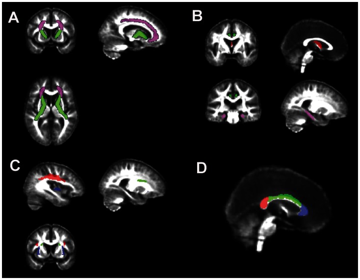
The goal of this study was to assess the relationship between Aβ deposition and white matter pathology (i.e., white matter hyperintensities, WMH) on microstructural integrity of the white matter. Fifty-seven participants (mean age: 78±7 years) from an ongoing multi-site research program who spanned the spectrum of normal to mild cognitive impairment (Clinical dementia rating 0-0.5) and low to high risk factors for arteriosclerosis and WMH pathology (defined as WMH volume >0.5% total intracranial volume) were assessed with positron emission tomography (PET) with Pittsburg compound B (PiB) and magnetic resonance and diffusion tensor imaging (DTI). Multivariate analysis of covariance were used to investigate the relationship between Aβ deposition and WMH pathology on fractional anisotropy (FA) from 9 tracts of interest (i.e., corona radiata, internal capsule, cingulum, parahippocampal white matter, corpus callosum, superior longitudinal, superior and inferior front-occipital fasciculi, and fornix). WMH pathology was associated with reduced FA in projection (i.e., internal capsule and corona radiate) and association (i.e., superior longitudinal, superior and inferior fronto-occipital fasciculi) fiber tracts. Aβ deposition (i.e., PiB positivity) was associated with reduced FA in the fornix and splenium of the corpus callosum. There were interactions between PiB and WMH pathology in the internal capsule and parahippocampal white matter, where Aβ deposition reduced FA more among subjects with WMH pathology than those without. However, accounting for apoE ε4 genotype rendered these interactions insignificant. Although this finding suggests that apoE4 may increase amyloid deposition, both in the parenchyma (resulting in PiB positivity) and in blood vessels (resulting in amyloid angiopathy and WMH pathology), and that these two factors together may be associated with compromised white matter microstructural integrity in multiple brain regions, additional studies with a longitudinal design will be necessary to resolve this issue.
Conflict of interest statement
Figures
Similar articles
-
Associations between white matter microstructure and amyloid burden in preclinical Alzheimer's disease: A multimodal imaging investigation.Neuroimage Clin. 2014 Feb 19;4:604-14. doi: 10.1016/j.nicl.2014.02.001. eCollection 2014. Neuroimage Clin. 2014. PMID: 24936411 Free PMC article.
-
Cerebral White Matter and Slow Gait: Contribution of Hyperintensities and Normal-appearing Parenchyma.J Gerontol A Biol Sci Med Sci. 2016 Jul;71(7):968-73. doi: 10.1093/gerona/glv224. Epub 2016 Jan 11. J Gerontol A Biol Sci Med Sci. 2016. PMID: 26755683 Free PMC article.
-
Impact of Apolipoprotein E4 Polymorphism on the Gray Matter Volume and the White Matter Integrity in Subjective Memory Impairment without White Matter Hyperintensities: Voxel-Based Morphometry and Tract-Based Spatial Statistics Study under 3-Tesla MRI.J Neuroimaging. 2016 Jan-Feb;26(1):144-9. doi: 10.1111/jon.12207. Epub 2015 Feb 11. J Neuroimaging. 2016. PMID: 25678236
-
Association between white matter alterations and domain-specific cognitive impairment in cerebral small vessel disease: A meta-analysis of diffusion tensor imaging.Front Aging Neurosci. 2022 Nov 22;14:1019088. doi: 10.3389/fnagi.2022.1019088. eCollection 2022. Front Aging Neurosci. 2022. PMID: 36483114 Free PMC article.
-
The application of diffusion tensor imaging in patients with mild cognitive impairment: a systematic review and meta-analysis.Front Neurol. 2025 Apr 23;16:1467578. doi: 10.3389/fneur.2025.1467578. eCollection 2025. Front Neurol. 2025. PMID: 40337171 Free PMC article.
Cited by
-
Review of 'the potential role of arterial stiffness in the pathogenesis of Alzheimer's disease'.Neurodegener Dis Manag. 2015;5(2):121-35. doi: 10.2217/nmt.14.53. Neurodegener Dis Manag. 2015. PMID: 25894876 Free PMC article. Review.
-
Independent value added by diffusion MRI for prediction of cognitive function in older adults.Neuroimage Clin. 2017 Jan 25;14:166-173. doi: 10.1016/j.nicl.2017.01.026. eCollection 2017. Neuroimage Clin. 2017. PMID: 28180075 Free PMC article.
-
Amyloid PET imaging: applications beyond Alzheimer's disease.Clin Transl Imaging. 2015;3(1):39-55. doi: 10.1007/s40336-014-0098-3. Epub 2015 Jan 21. Clin Transl Imaging. 2015. PMID: 25741489 Free PMC article. Review.
-
White matter hyperintensities, cognitive impairment and dementia: an update.Nat Rev Neurol. 2015 Mar;11(3):157-65. doi: 10.1038/nrneurol.2015.10. Epub 2015 Feb 17. Nat Rev Neurol. 2015. PMID: 25686760 Review.
-
Characterization of white matter changes along fibers by automated fiber quantification in the early stages of Alzheimer's disease.Neuroimage Clin. 2019;22:101723. doi: 10.1016/j.nicl.2019.101723. Epub 2019 Feb 18. Neuroimage Clin. 2019. PMID: 30798166 Free PMC article.
References
-
- Neuropathology Group of the Medical Research Council Cognitive Function and Aging Study (MRC CFAS) (2001) Pathological correlates of late-onset dementia in a multicentre, community-based population in England and Wales. Lancet 357: 169–175. - PubMed
-
- Breteler MM, van Swieten JC, Bots ML, Grobbee DE, Claus JJ, et al. (1994) Cerebral white matter lesions, vascular risk factors, and cognitive function in a population-based study: the Rotterdam Study. Neurology 44: 1246–1252. - PubMed
-
- de Leeuw FE, de Groot JC, Oudkerk M, Witteman JC, Hofman A, et al. (200) Aortic atherosclerosis at middle age predicts cerebral white matter lesions in the elderly. Stroke 31: 425–429. - PubMed
-
- de Leeuw FE, de Groot JC, Oudkerk M, Witteman JC, Hofman A, et al. (2002) Hypertension and cerebral white matter lesions in a prospective cohort study. Brain 125: 765–772. - PubMed
Publication types
MeSH terms
Substances
Grants and funding
LinkOut - more resources
Full Text Sources
Other Literature Sources
Medical
Miscellaneous
