. 2013 Jun 7;8(6):e65384.
doi: 10.1371/journal.pone.0065384. Print 2013.

Structurally designed attenuated subunit vaccines for S. aureus LukS-PV and LukF-PV confer protection in a mouse bacteremia model

Affiliations

Structurally designed attenuated subunit vaccines for S. aureus LukS-PV and LukF-PV confer protection in a mouse bacteremia model

Hatice Karauzum et al. PLoS One. .

Abstract

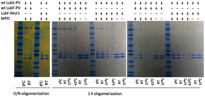
Previous efforts towards S. aureus vaccine development have largely focused on cell surface antigens to induce opsonophagocytic killing aimed at providing sterile immunity, a concept successfully applied to other Gram-positive pathogens such as Streptococcus pneumoniae. However, these approaches have largely failed, possibly in part due to the remarkable diversity of the staphylococcal virulence factors such as secreted immunosuppressive and tissue destructive toxins. S. aureus produces several pore-forming toxins including the single subunit alpha hemolysin as well as bicomponent leukotoxins such as Panton-Valentine leukocidin (PVL), gamma hemolysins (Hlg), and LukED. Here we report the generation of highly attenuated mutants of PVL subunits LukS-PV and LukF-PV that were rationally designed, based on an octameric structural model of the toxin, to be deficient in oligomerization. The attenuated subunit vaccines were highly immunogenic and showed significant protection in a mouse model of S. aureus USA300 sepsis. Protection against sepsis was also demonstrated by passive transfer of rabbit immunoglobulin raised against LukS-PV. Antibodies to LukS-PV inhibited the homologous oligomerization of LukS-PV with LukF-PV as well heterologous oligomerization with HlgB. Importantly, immune sera from mice vaccinated with the LukS mutant not only inhibited the PMN lytic activity produced by the PVL-positive USA300 but also blocked PMN lysis induced by supernatants of PVL-negative strains suggesting a broad protective activity towards other bicomponent toxins. These findings strongly support the novel concept of an anti-virulence, toxin-based vaccine intended for prevention of clinical S. aureus invasive disease, rather than achieving sterile immunity. Such a multivalent vaccine may include attenuated leukotoxins, alpha hemolysin, and superantigens.


Conflict of interest statement

Competing Interests: The authors have read the journal’s policy and have the following conflicts: Tam Nguyen served as a consultant and all other authors were employed by Integrated Biotherapeutics, Inc. at the time work was performed. M. Javad Aman and Kelly Warfield are shareholders in the company of the Inegrated BioTherapeutics Inc., the organization at which this work was performed. Drs. Chris Clegg and Paul Sleath (Immune Design Corp.) provided GLA-SE adjuvant and NARSA repository for USA300 and NCTC8325-4 (NRS77) for this study. The authors have filed a patent application for the described mutants. This patent is not published yet but is expected to be published in July 2013. The pending application number is PCT/US12/67483. The title of invention is IMMUNOGENIC COMPOSITION COMPRISING PANTON-VALENTINE LEUKOCIDIN (PVL) DERIVED POLYPEPTIDES. There are no further patents, products in development or marketed products to declare. This does not alter the authors’ adherence to all the PLOS ONE policies on sharing data and materials, as detailed online in the guide for authors.

Figures

Figure 1
Figure 1. Key residues involved in interaction of LukS-PV and LukF-PV.
A) Interface interactions between T28 of LukS-PV (green ribbon) and N158 and F159 of LukF-PV (pink ribbon). B) Interface interaction between S209 of LukS-PV (green ribbon) and K102 of LukF-PV (pink ribbon).
Figure 2
Figure 2. SDS-PAGE and Western blot of mutant and wild type forms of LukS-PV and LukF-PV.
A) LukS-PV wt and mutants 2–9, B) LukF-PV wt and mutants 1–3. Upper panels: coomassie staining; Lower panels: Western blot.
Figure 3
Figure 3. Attenuation of PMN lytic activity of PVL mutants.
A) % survival of HL-60 derived neutrophils treated with increasing concentrations of wt or mutant LukS-PV each in combination with wt LukF-PV. Results represent mean values from 5 independent experiments. STDV is shown only for wt and the triple mutant (LukS-Mut9). B) % survival of HL-60 derived neutrophils treated with increasing concentrations of wt or mutant LukF-PV each in combination with wt LukS-PV or LukS-PV triple mutant (LukS-Mut9).
Figure 4
Figure 4. Thermal unfolding of LukS-PV and LukF-PV proteins as monitored by thermofluor assay using Sypro Orange dye.
A) Plot of fluorescence intensity of PVL proteins at 588 nm against temperature. Data was collected for every 5°C. B) Plot of fraction unfolded calculated from the thermal denaturation curve. Key: LukS-PV wild type, LukS-PV T28F/S209A (LukS-Mut8), LukS-PV T28F/K97A/S209A (LukS-Mut9), LukF-PV Wild type and LukF-PV K102A (LukF-Mut1) were tested.
Figure 5
Figure 5. Homologous and heterologous oligomerization of wt and mutant LukS-PV and LukF-PV.
Individual subunits as indicated above the panel were co-incubated overnight at a concentration of 30 µg/ml with 40%MPD and analyzed by SDS-PAGE without boiling. Lane 1: molecular weight Marker; Lanes 2–5: different combinations of wild type (wt) or mutant leukocidins as shown above the panel.
Figure 6
Figure 6. LukF-Mut1 displays reduced oligomerization activity.
Individual subunits as indicated above the panel were co-incubated with or without MPD at a concentration of 30 µg/ml(Left and right panels) or 100 µg/ml (middle panel) at indicated temperatures and analyzed by SDS-PAGE without boiling. Incubation time was either 24 hours (left panel) or 1 hour (middle and right panels). RT: room temperature.
Figure 7
Figure 7. Immunogenicity of LukS-Mut9 with different adjuvants in mice.
A) Total Ab titers determined by ELISA for individual mouse sera (EC50; i.e. dilution of serum with 50% maximal signal on ELISA plates coated with wild type LukS-PV). B) Neutralization determined in HL-60 toxin neutralization assay using wild type LukS-PV and LukF-PV toxins. Percent neutralization of wild type toxin is shown at 1∶100 dilution of serum from vaccinated mice (sera pooled from 5 mice in each group). Doses used: antigens: 10 ug; Al(OH)3∶34 µg, AlPO4∶70 µg, GLA-SE: 20 ug, and CpG: 10 µg/mouse.
Figure 8
Figure 8. Imunogenicity and protective efficacy of LukS-Mut9 and LukF-Mut1.
A) Antibody titers of mice immunized with lukS-Mut9 or LukF-Mut1 towards the homologous wild type antigens or the combination of both mutants towards each antigen. B) Protection from lethal challenge by active immunization with LukS-Mut9 and LukF-Mut1 along with GLA-SE in USA300 bacteremia model.
Figure 9
Figure 9. Effect of active immunization with PVL mutants on bacterial load.
Bacterial loads were determined in blood and various organs of immunized mice 3 days post challenge with a sub-lethal dose of USA300.
Figure 10
Figure 10. Inhibition of oligomerization by anti LukS-PV polyclonal antibody.
A) Lane1: Marker; lanes 2–8:, 30 µg/ml of LukS-PV was incubated for 30 min at RT with anti-LukS-PV rabbit polyclonal antibodies (pAbs) at 2-fold decreasing concentrations (1.1 mg/ml to 0.017 mg/ml) and then equal concentration of LukF-PV subunit was added; lane 9: LukS+LukF without pAbs; lane10: LukS+LukF+pAbs without MPD; lane 11: pAbs+MPD only. B) Inhibition of oligomeric band formed by LukS-PV+hlgB by anti-LukS-PV pAb. Lane1: Marker; lanes 2–8∶30 ug/ml of LukS-PV was incubated for 30 min at RT with anti-LukS-PV rabbit polyclonal antibodies (pAbs) at 2-fold decreasing concentrations (1.1 mg/ml to 0.017 mg/ml) then equal concentration of HlgB subunit was added; lane 9: LukS-PV+HlgB without pAbs; lane10: LukS-PV+HlgB+pAbs without MPD; lanes 11–14 LukS-PV+HlgB along with naïve rabbit IgG (1.1 mg/ml to 0.14 mg/ml), and lane 15: rabbit anti-LukS-PV pAbs +MPD only.
Figure 11
Figure 11. LukS-Mut9 induces cross reactive antibodies.
Serum samples after 4 immunizations with LukS-Mut9 were tested for ELISA titers against WT LukS-PV (A) and HlgC (B).
Figure 12
Figure 12. LukS-Mut9 antisera cross neutralize PMN lytic activity induced by other leukocidins.
Inhibition by LukS-Mut 9 immunized sera of PMN lytic activity induced by S. aureus strain 8325-4 (PVL-neg) supernatant (A), USA300 (PVL-pos) Supernatant (B), purified PVL (S+F subunits) (C), and purified Hlg (B+C subunits) (D).

References

    1. Nizet V (2007) Understanding how leading bacterial pathogens subvert innate immunity to reveal novel therapeutic targets. J Allergy Clin Immunol 120: 13–22. - PubMed
    1. Kotzin BL, Leung DY, Kappler J, Marrack P (1993) Superantigens and their potential role in human disease. Adv Immunol 54: 99–166. - PubMed
    1. Meyer RD, Monday SR, Bohach GA, Schlievert SM (2001) Prolonged course of toxic shock syndrome associated with methicillin-resistant Staphylococcus aureus enterotoxins G and I. Int J Infect Dis. 5: 163–166. - PubMed
    1. Schuberth HJ, Krueger C, Zerbe H, Bleckmann E, Leibold W (2001) Characterization of leukocytotoxic and superantigen-like factors produced by Staphylococcus aureus isolates from milk of cows with mastitis. Vet Microbiol 82: 187–199. - PubMed
    1. Tristan A, Ferry T, Durand G, Dauwalder O, Bes M, et al. (2007) Virulence determinants in community and hospital meticillin-resistant Staphylococcus aureus. J Hosp Infect 65 Suppl 2105–109. - PubMed

Publication types

MeSH terms