. 2013 Jun 7;8(6):e65454.
doi: 10.1371/journal.pone.0065454. Print 2013.

A Saccharomyces cerevisiae assay system to investigate ligand/AdipoR1 interactions that lead to cellular signaling

Affiliations

A Saccharomyces cerevisiae assay system to investigate ligand/AdipoR1 interactions that lead to cellular signaling

Mustapha Aouida et al. PLoS One. .

Abstract

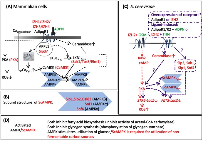
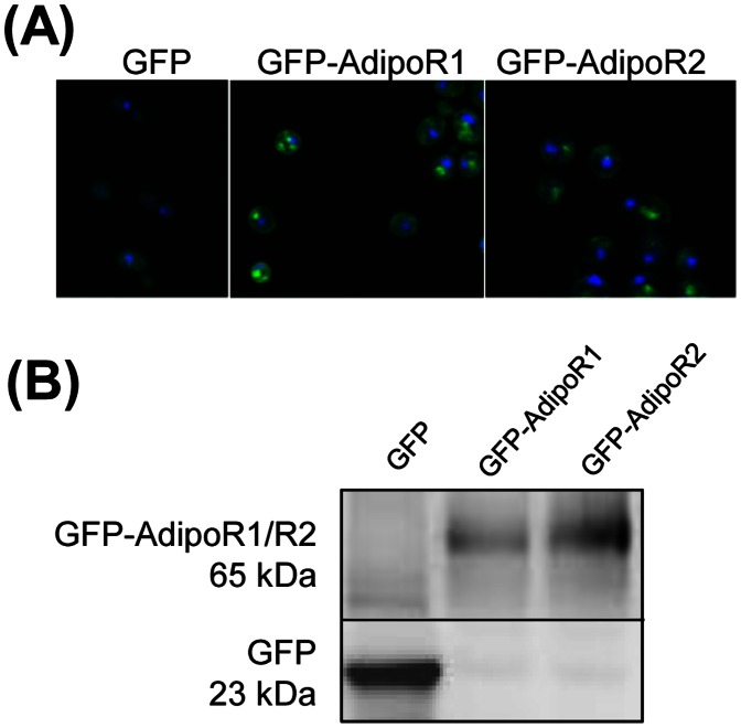
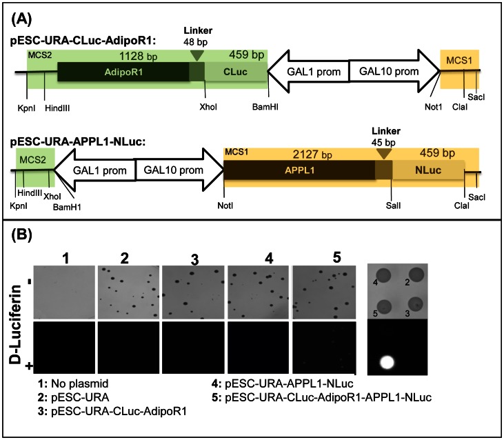
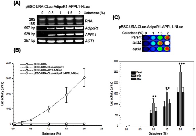
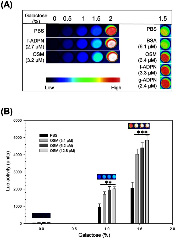
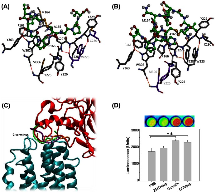
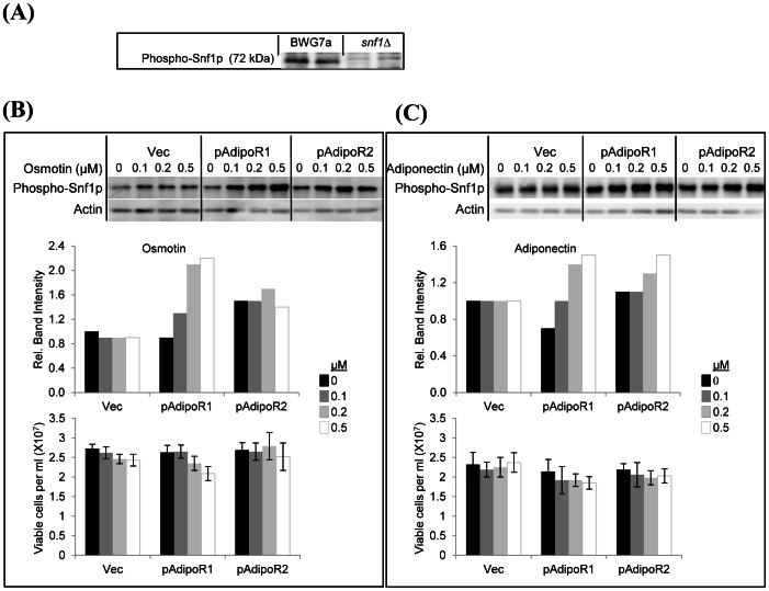
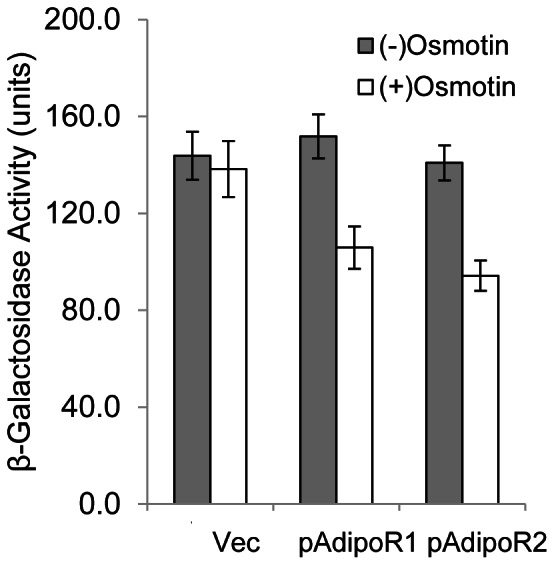
Adiponectin is a mammalian hormone that exerts anti-diabetic, anti-cancer and cardioprotective effects through interaction with its major ubiquitously expressed plasma membrane localized receptors, AdipoR1 and AdipoR2. Here, we report a Saccharomyces cerevisiae based method for investigating agonist-AdipoR interactions that is amenable for high-throughput scale-up and can be used to study both AdipoRs separately. Agonist-AdipoR1 interactions are detected using a split firefly luciferase assay based on reconstitution of firefly luciferase (Luc) activity due to juxtaposition of its N- and C-terminal fragments, NLuc and CLuc, by ligand induced interaction of the chimeric proteins CLuc-AdipoR1 and APPL1-NLuc (adaptor protein containing pleckstrin homology domain, phosphotyrosine binding domain and leucine zipper motif 1-NLuc) in a S. cerevisiae strain lacking the yeast homolog of AdipoRs (Izh2p). The assay monitors the earliest known step in the adiponectin-AdipoR anti-diabetic signaling cascade. We demonstrate that reconstituted Luc activity can be detected in colonies or cells using a CCD camera and quantified in cell suspensions using a microplate reader. AdipoR1-APPL1 interaction occurs in absence of ligand but can be stimulated specifically by agonists such as adiponectin and the tobacco protein osmotin that was shown to have AdipoR-dependent adiponectin-like biological activity in mammalian cells. To further validate this assay, we have modeled the three dimensional structures of receptor-ligand complexes of membrane-embedded AdipoR1 with cyclic peptides derived from osmotin or osmotin-like plant proteins. We demonstrate that the calculated AdipoR1-peptide binding energies correlate with the peptides' ability to behave as AdipoR1 agonists in the split luciferase assay. Further, we demonstrate agonist-AdipoR dependent activation of protein kinase A (PKA) signaling and AMP activated protein kinase (AMPK) phosphorylation in S. cerevisiae, which are homologous to important mammalian adiponectin-AdipoR1 signaling pathways. This system should facilitate the development of therapeutic inventions targeting adiponectin and/or AdipoR physiology.


Conflict of interest statement

Competing Interests: The authors declare the affiliation of MLN to Bio-Write, LLC, does not represent a conflict of interest in the presentation, peer review, editorial decision-making, or publication of this manuscript. This does not alter the authors’ adherence to all the PLOS ONE policies on sharing data and materials. None of the other authors have potential competing interests. There are no patent applications or products in development or marketed products based on the results in this paper.

Figures

Figure 1
Figure 1. Summary of AdipoR-mediated signaling pathways in mammalian cells and IZH2-mediated signaling pathways in S. cerevisiae cells.
(A) The components of the AdipoR1 signaling pathway of mammalian cells are shown in black font and their S. cerevisiae homologs, if known are shown in red font. In mammalian cells, adaptor protein containing pleckstrin homology domain, phosphotyrosine binding domain and leucine zipper motif 1 (APPL1) interacts directly with AdipoR1. Interaction of adiponectin (ADPN) with AdipoR1 stimulates AdipoR1-APPL1 interaction. This results in release of Ca2+ from the ER to the cytosol and also increases export of LKB1 kinase from the nucleus. Influx of extracellular Ca2+ to the cytosol is also stimulated by adiponectin, although the mechanism by which this occurs remains to be clarified. Increase in cytosolic Ca2+ concentration activates Ca2+/calmodulin-dependent protein kinase kinase (CaMKK) which in turn activates AMP activated protein kinase (AMPK) by phosphorylating its α subunit. However, phosphorylation of AMPK α subunit by the cytosol-localized kinase LKB1 is the major pathway for activation of AMPK. Adiponectin-AdipoR1 interaction also increases cellular ceramidase activity which in turn leads to phosphorylation of AMPKα subunit. The details of this pathway are not clear yet. Activation of AMPK is required for many of the anti-diabetic and anti-atherosclerotic effects of adiponectin. In vascular endothelial cells, interaction of AdipoR1 with adiponectin activates protein kinase A (PKA) which has the effect of lowering accumulation of reactive oxygen species (ROS) and thereby reducing inflammation. (B) Subunit structure of S. cerevisiae AMPK (ScAMPK). Like mammalian AMPK, ScAMPK is a trimer composed of an α, β, and γ subunit. The genes encoding the β subunit isoforms as well as the sole α and γ subunits are indicated. (C) Components of the IZH2-mediated signaling pathways in S. cerevisiae are shown in red font. Interaction of IZH2 (homolog of AdipoRs) with osmotin (OSM) activates PKA via a RAS2-cAMP pathway. Overexpression of IZH2, overexpression of AdipoR1, treatment of S. cerevisiae cells expressing AdipoR1 with adiponectin and treatment of S. cerevisiae cells expressing various levels of IZH2 with thaumatin (THN, a homolog of osmotin) has been shown to activate PKA by increasing cellular ceramidase activity. Activation of PKA leads to decreased transcription from a stress responsive promoter element (STRE) and increased cellular ROS content. Activated PKA promotes export of ScAMPK from the nucleus which leads to decreased transcription from the ferroxidase (FET3) promoter. Activated PKA also represses FET3 transcription via the stress-responsive transcription factors MSN2/4. Mutational analyses show that genes encoding the APPL1-lke protein Sip3, the LKB1-like protein Sak1, the ScAMPK β subunit Sip1 and the ScAMPK γ subunit Snf4 are components of the pathway leading from IZH2 (or AdipoR1) to FET3 repression in S. cerevisiae.
Figure 2
Figure 2. AdipoRs are expressed on the plasma membrane in S. cerevisiae.
(A) Subcellular localization of AdipoRs by confocal microscopy. S. cerevisiae strain BY4741 carrying plasmid pYES-EGFP (GFP), pYES-EGFP-AdipoR1 (GFP-AdipoR1) and pYES-EGFP-AdipoR2 (GFP-AdipoR2) were cultured in selective minimal medium containing 2% galactose. Shown are images of cells that were in the early log phase of growth. (B) Western blot analysis of total membrane protein extracts that were fractionated by 10% SDS-PAGE. The predicted molecular weights of GFP-AdipoR1 and GFP-AdipoR2 are around 65 kDa.
Figure 3
Figure 3. The split firefly luciferase complementation colony assay for AdipoR1 and APPL1 interaction.
(A) Schematic representation of the plasmid vector constructs. CLuc-AdipoR1 was cloned into the multiple cloning site 2 (MCS2, green shadow) of pESC-URA for expression from the GAL1 promoter and Appl1-NLuc was cloned into MCS1 (orange shadow) of pESC-URA for expression from the GAL10 promoter. pESC-URA-CLuc-AdipoR1-APPL1-NLuc (not shown) contains the depicted APPL1-NLuc construct inserted between Not1 and Sac1 sites of the MCS1 of pESC-URA-CLuc-AdipoR1. (B) Visualization of the AdipoR1-APPL1 interaction in colonies. Cells of strain BY4741 untransformed (No plasmid) or transformed with the indicated plasmids were spread on minimal medium lacking uracil containing 2% galactose. After 2 days incubation at 30°C, plates were photographed before (–) and imaged after (+) spraying with D-luciferin.
Figure 4
Figure 4. Luciferase reporter activity depends on the expression level of AdipoR1 and APPL1.
(A) RT-PCR analysis of AdipoR1 and APPL1 expression in total RNA (2 µg) from cells of strain BY4741 carrying pESC-URA-CLuc-AdipoR1-APPL1-NLuc that were grown for 16 h at 30°C in selective minimal medium at the indicated galactose concentrations. The final concentration of sugars in the growth media was adjusted to 2% with raffinose. ACT1 expression is shown for normalization. (B) Quantification of luciferase activity in BY4741 cells carrying pESC-URA, pESC-URA-CLuc-AdipoR1, pESC-URA-APPL1-NLuc and pESC-URA-CLuc-AdipoR1-APPL1-NLuc plasmids. Cells were grown for 16 h at 30°C in selective minimal medium at the indicated galactose concentrations. Luciferase activity measurements were made as described in Methods. (C) Imaging (top) and quantification of luciferase activity (bottom) in pESC-URA-CLuc-AdipoR1-APPL1-NLuc transformants of strain BY4741 (Parent) or its isogenic izh2Δ and sip3Δ derivative strains. Assays were performed as described above on cells that were grown for 16 h at 30°C in selective minimal medium at the indicated galactose concentrations. Results are expressed as the mean ± SD from three separate experiments with triplicate samples. Significant difference from the parent strain is indicated by asterisks. Symbols: **, p<0.01; ***, p<0.001.
Figure 5
Figure 5. The AdipoR1 ligands, adiponectin and osmotin, induce increase in Luc reporter activity.
Cells of strain BY4741carrying pESC-URA-CLuc-AdipoR1-APPL1-NLuc were grown for 16 h at 30°C in selective minimal medium at the indicated galactose concentrations, treated for 4 h at 30°C with the indicated test compounds and then assayed for Luc activity. (A) Imaging of Luc activity. (B) Quantitative measurement of Luc activity as a function of osmotin concentration. A representative image of relative Luc activity at the different osmotin concentrations is shown for each galactose concentration. Data represent the means ± SD from three experiments with triplicate samples. For each galactose concentration, significant differences by a Student’s t-test between osmotin treated samples and untreated control are indicated by asterisks. Symbols: PBS, 1/8 X PBS; f-ADPN, bacterially expressed full length adiponectin; BSA, bovine serum albumin, OSM, osmotin; g-ADPN, bacterially expressed globular adiponectin; **, p<0.01; ***, p<0.001.
Figure 6
Figure 6. Comparison of TLP-derived peptide ligands binding to AdipoR1.
3D model of (A) OSMpep and (B) ZMTNpep bound to AdipoR1. Peptides are shown in ball and stick representation (C atoms in green). The interacting residues of AdipoR1 are shown as sticks only (interacting residues: C in gray; non-interacting residues: C in purple). Atom color coding is white = H, red = O, blue = N, yellow = S. (C) Overlay of osmotin (red) and the three top scoring OSMpep poses (peptide strands in green, purple and orange illustrate orientation of poses with 1st, 2nd and 3rd highest docking score) bound to AdipoR1 (blue). (D) Verification of interaction strength of the peptides with AdipoR1 by the split luciferase assay. Cells of strain BY4741carrying pESC-URA-CLuc-AdipoR1-APPL1-NLuc were grown for 16 h at 30°C in selective minimal medium containing 1.5% galactose, treated for 4 h at 30°C with the indicated test compounds and then used for luciferase activity measurement. The concentrations of test compounds in the assay were: PBS, 1/8 X PBS; ZMTNpep, 80 µg/mL; osmotin, 80 µg/mL (3.1 µM) and OSMpep, 80 µg/mL. Shown is a representative image of luciferase activity and quantification of the luminescence under the CCD camera. Data are the mean ±SD from three separate experiments with triplicate samples. Asterisks represent significant differences at **, p<0.01 by Student’s t-test.
Figure 7
Figure 7. Osmotin signaling mediated by adiponectin receptors induces Snf1p phosphorylation in an izh2Δ mutant.
(A) Cell lysates (100 µg protein) of strain BWG1-7a and an isogenic Δsnf1::Kan_MX line were fractionated by 10% SDS-PAGE. Shown are blots probed with anti-phospho-AMPK(Thr-172) antibody. The expected size of Snf1p is 72 kDa. (B, C) Cells (about 108/mL) of strain BWG1-7a Δizh2::Kan_MX transformed with p426GPD (Vec), p426GPD-AdipoR1 (pAdipoR1) or p426GPD-AdipoR2 (pAdipoR2) were treated with indicated osmotin and adiponectin concentrations for 30 min at 30°C in YPD. Aliquots were withdrawn for viable counts determination before the cell lysates were prepared for analysis by10% SDS-PAGE. Shown in the top panels are blots probed first with phospho-AMPK(Thr-172) antibody, then stripped and probed with actin antibody (100 µg total protein per lane). Shown in the middle panels are relative band intensities in the depicted gels. ‘Relative band intensity’ was defined as the ratio of the intensity of phospho-Snf1p signal to actin signal for each lane when the value of this ratio for the corresponding untreated Vec sample was arbitrarily assigned the value 1.0. Shown in the bottom panels are viable counts in each sample at the end of the treatments. The experiments were performed twice with comparable results and the results of one experiment are shown.
Figure 8
Figure 8. Osmotin signaling mediated by adiponectin receptors induces suppression of gene expression via stress responsive promoter elements.
Cells (about 108/mL) of strain BWG1-7a Δizh2::Kan_MX co-transformed with pSTRE-lacZ(LEU2) and p426-GPD (Vec), p426-GPD-AdipoR1 (pAdipoR1) or p426-GPD-AdipoR2 (pAdipoR2) were treated with osmotin (4 µM) in YPD for 45 min at 30°C and β-galactosidase activity was measured at the end of the treatment period. The values represent the means ± SE of two independent experiments with 3 to 4 samples per experiment.

References

    1. Berg AH, Combs TP, Scherer PE (2002) ACRP30/adiponectin: an adipokine regulating glucose and lipid metabolism. Trends Endocrinol Metab 13: 84–89. - PubMed
    1. Jortay J, Senou M, Abou-Samra M, Noel L, Robert A, et al. (2012) Adiponectin and skeletal muscle: pathophysiological implications in metabolic stress. Am J Pathol 181: 245–256. - PubMed
    1. Kadowaki T, Yamauchi T (2005) Adiponectin and adiponectin receptors. Endocr Rev 26: 439–451. - PubMed
    1. Wang Y, Lau WB, Gao E, Tao L, Yuan Y, et al. (2010) Cardiomyocyte-derived adiponectin is biologically active in protecting against myocardial ischemia-reperfusion injury. Am J Physiol Endocrinol Metab 298: E663–670. - PMC - PubMed
    1. Hara K, Boutin P, Mori Y, Tobe K, Dina C, et al. (2002) Genetic variation in the gene encoding adiponectin is associated with an increased risk of type 2 diabetes in the Japanese population. Diabetes 51: 536–540. - PubMed

Publication types

MeSH terms