

Cone beam computed tomography evaluation of the diagnosis, treatment planning, and long-term followup of large periapical lesions treated by endodontic surgery: two case reports
- PMID: 23762646
- PMCID: PMC3674742
- DOI: 10.1155/2013/564392
Cone beam computed tomography evaluation of the diagnosis, treatment planning, and long-term followup of large periapical lesions treated by endodontic surgery: two case reports
Abstract
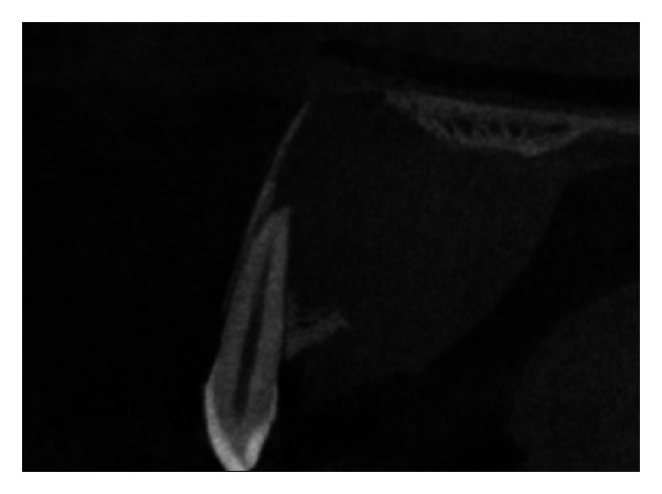
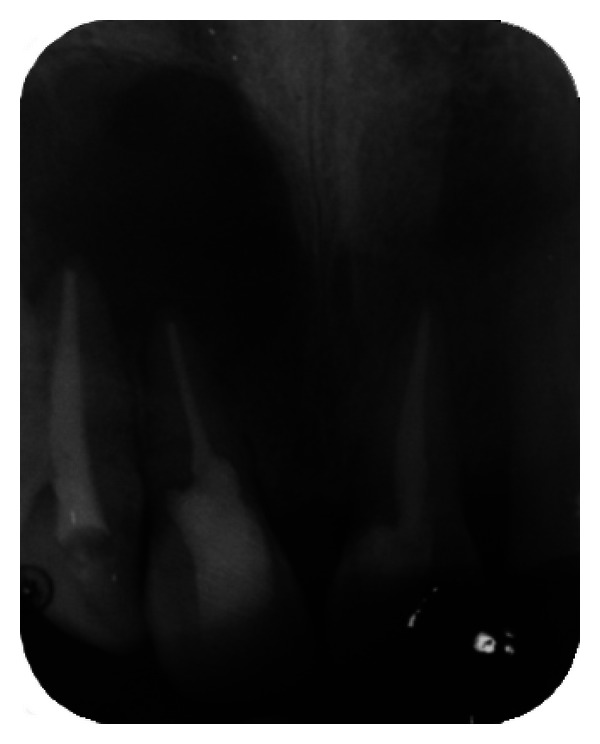
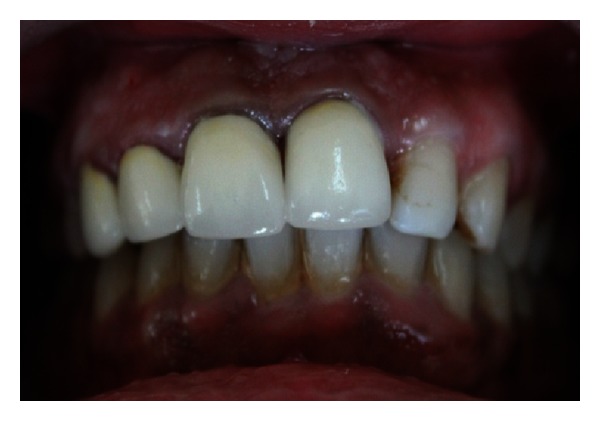
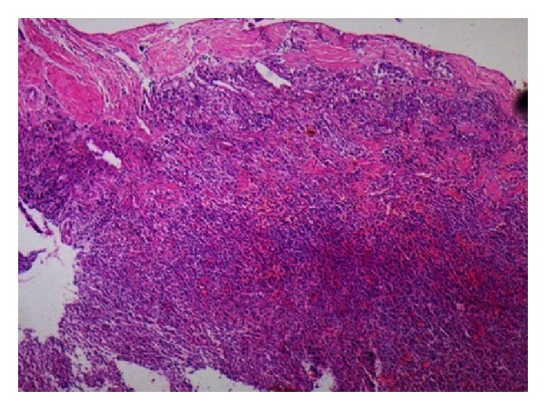
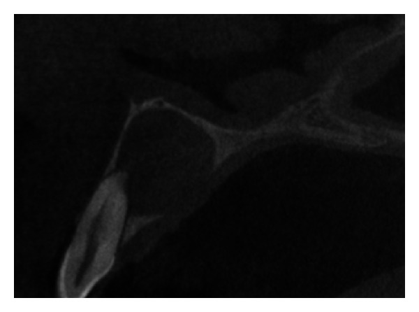
The aim of this case report is to present two cases where cone beam computed tomography (CBCT) was used for the diagnosis, treatment planning, and followup of large periapical lesions in relation to maxillary anterior teeth treated by endodontic surgery. Periapical disease may be detected sooner using CBCT, and their true size, extent, nature, and position can be assessed. It allows clinician to select the most relevant views of the area of interest resulting in improved detection of periapical lesions. CBCT scan may provide a better, more accurate, and faster method to differentially diagnose a solid (granuloma) from a fluid-filled lesion or cavity (cyst). In the present case report, endodontic treatment was performed for both the cases followed by endodontic surgery. Biopsy was done to establish the confirmatory histopathological diagnosis of the periapical lesions. Long-term assessment of the periapical healing following surgery was done in all the three dimensions using CBCT and was found to be more accurate than IOPA radiography. It was concluded that CBCT was a useful modality in making the diagnosis and treatment plan and assessing the outcome of endodontic surgery for large periapical lesions.
Figures


References
-
- Estrela C, Bueno MR, Leles CR, Azevedo B, Azevedo JR. Accuracy of cone beam computed tomography and panoramic and periapical radiography for detection of apical periodontitis. Journal of Endodontics. 2008;34(3):273–279. - PubMed
-
- de Paula-Silva FWG, Júnior MS, Leonardo MR, Consolaro A, da Silva LAB. Cone-beam computerized tomographic, radiographic, and histologic evaluation of periapical repair in dogs’ post-endodontic treatment. Oral Surgery, Oral Medicine, Oral Pathology, Oral Radiology and Endodontology. 2009;108(5):796–805. - PubMed
-
- Simon JHS, Enciso R, Malfaz JM, Roges R, Bailey-Perry M, Patel A. Differential diagnosis of large periapical lesions using cone-beam computed tomography measurements and biopsy. Journal of Endodontics. 2006;32(9):833–837. - PubMed
-
- Patel S, Dawood A, Pitt Ford T, Whaites E. The potential applications of cone beam computed tomography in the management of endodontic problems. International Endodontic Journal. 2007;40(10):818–830. - PubMed
LinkOut - more resources
Full Text Sources
Other Literature Sources
