

Giant synovial cyst of thigh: a rare entity
- PMID: 23762703
- PMCID: PMC3670511
- DOI: 10.1155/2013/967215
Giant synovial cyst of thigh: a rare entity
Abstract
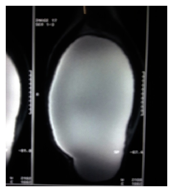
Synovial cyst occurs secondary to traumatic, degenerative, or inflammatory conditions. Synovial cysts represent abnormal distension of bursae, which communicate with the joint. Giant synovial cysts are typically due to rheumatoid arthritis, other causes being trauma and synovial pseudoarthrosis. A 33-year-old male presented to an outpatient clinic with a massive swelling on his posterolateral aspect of right thigh extending from upper one-third to the knee joint which had been increasing in size over the past six months. This was associated with dull aching pain. All laboratory investigations were within normal parameters. Even FNAC was inconclusive. With time, swelling was increasing in size. Ultrasound revealed the cystic nature of swelling. MRI showed large cystic lesion 24 × 10 × 12 cm in posterolateral aspect of thigh extending up to knee joint. Following the MRI, an excision was planned. After excision, histological examination confirmed the synovial nature of the cyst, which had a collagenous wall and dense chronic inflammatory cells. As the disease is extremely rare and asymptomatic, precise diagnosis is difficult and often delayed. We consider that open surgical excision should be reserved for cases of large synovial cysts because it can provide a complete resection of the lesion and minimize the risk of recurrence.
Figures


Similar articles
-
Giant synovial cyst of knee treated arthroscopically through a cystic portal.Knee Surg Sports Traumatol Arthrosc. 2008 Feb;16(2):175-8. doi: 10.1007/s00167-007-0405-x. Epub 2007 Sep 26. Knee Surg Sports Traumatol Arthrosc. 2008. PMID: 17899006
-
Scalloping Sacral Arachnoid Cyst as a Cause of Perianal Pain- A Case Report.J Orthop Case Rep. 2014 Apr-Jun;4(2):28-32. doi: 10.13107/jocr.2250-0685.163. J Orthop Case Rep. 2014. PMID: 27298955 Free PMC article.
-
Treatment of temporomandibular joint ganglion cyst.J Craniofac Surg. 2011 Sep;22(5):1935-7. doi: 10.1097/SCS.0b013e318211516d. J Craniofac Surg. 2011. PMID: 21959472
-
Cysts about the knee: evaluation and management.J Am Acad Orthop Surg. 2013 Aug;21(8):469-79. doi: 10.5435/JAAOS-21-08-469. J Am Acad Orthop Surg. 2013. PMID: 23908253 Review.
-
Synovial cyst of the temporomandibular joint: a case report and literature review.Int J Oral Maxillofac Surg. 2011 Aug;40(8):874-7. doi: 10.1016/j.ijom.2011.02.027. Epub 2011 Apr 5. Int J Oral Maxillofac Surg. 2011. PMID: 21470821 Review.
Cited by
-
Giant Intraosseous Cyst-Like Lesions of the Metacarpal Bones in Rheumatoid Arthritis.J Imaging. 2021 Jul 12;7(7):113. doi: 10.3390/jimaging7070113. J Imaging. 2021. PMID: 39080901 Free PMC article.
-
Endoscopic Resection of Lateral Synovial Cyst of the Knee.Arthrosc Tech. 2015 Dec 14;4(6):e815-8. doi: 10.1016/j.eats.2015.08.001. eCollection 2015 Dec. Arthrosc Tech. 2015. PMID: 27284517 Free PMC article.
-
Ganglion cyst versus synovial cyst? Ultrasound characteristics through a review of the literature.Rheumatol Int. 2015 Apr;35(4):597-605. doi: 10.1007/s00296-014-3120-1. Epub 2014 Sep 5. Rheumatol Int. 2015. PMID: 25190552 Review.
References
-
- Perri JA, Rodnan GP, Mankin HJ. Giant synovial cysts of the calf in patients with rheumatoid arthritis. Journal of Bone and Joint Surgery—Series A. 1968;50(4):709–719. - PubMed
-
- Pastershank SP, Mitchell DM. Knee joint bursal abnormalities in rheumatoid arthritis. Canadian Association of Radiologists Journal. 1977;28(3):199–203. - PubMed
-
- White TK, Incavo SJ, Moreland MS. Giant synovial cyst of the hip joint. Orthopaedic Review. 1988;17(6):609–612. - PubMed
-
- Lu KH. Unusual solitary ganglion cysts of the anterior segment of the lateral meniscus. Arthroscopy. 2003;19(3, article E16) - PubMed
-
- Shetty GM, Nha KW, Patil SP, et al. Ganglion cysts of the posterior cruciate ligament. Knee. 2008;15(4):325–329. - PubMed
LinkOut - more resources
Full Text Sources
Other Literature Sources
Research Materials
Miscellaneous
