. 2013 Mar 31:2013:167407.
doi: 10.1155/2013/167407.

Experimental Rat and Mouse Carotid Artery Surgery: Injury & Remodeling Studies

Affiliations

Experimental Rat and Mouse Carotid Artery Surgery: Injury & Remodeling Studies

Andrew W Holt et al. ISRN Minim Invasive Surg. .

Abstract

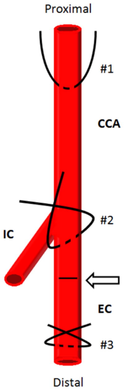
In cardiovascular research, translation of benchtop findings to the whole body environment is often critical in order to gain a more thorough and comprehensive clinical evaluation of the data with direct extrapolation to the human condition. In particular, developmental and/or pathophysiologic vascular growth studies often employ in vitro approaches such as cultured cells or tissue explant models in order to analyze specific cellular, molecular, genetic and/or biochemical signaling factors under pristine controlled conditions. However, validation of in vitro data in a whole body setting complete with neural, endocrine and other systemic contributions provides essential proof-of-concept from a clinical perspective. Several well-characterized experimental in vivo models exist that provide excellent proof-of-concept tools with which to examine vascular growth and remodeling in the whole body. This article will examine the rat carotid artery balloon injury model, the mouse carotid artery wire denudation injury model, and rat and mouse carotid artery ligation models with particular emphasis on minimally invasive surgical access to the site of intervention. Discussion will include key scientific and technical details as well as caveats, limitations, and considerations for practical use for each of these valuable experimental models.

Keywords: balloon injury; carotid artery; ligation; mouse; neointima; rat; remodeling; vascular endothelial cell; vascular smooth muscle cell; wire denudation.


Figures

Figure 1
Figure 1
Simplified schematic showing carotid artery vasculature and sites for placement of arterial sutures for retraction and/or ligation. During vascular access suture #1 is looped around the most proximal portion of the left common carotid artery (CCA), suture #2 is looped around the external branch immediately distal to the internal carotid (IC) and external carotid (EC) bifurcation, and suture #3 is looped around the EC artery as distally as possible. The arteriotomy site for insertion of the balloon catheter is shown on the EC branch between the bifurcation and the site of distal ligature (#3) and is indicated by an arrow and dashed line.
Figure 2
Figure 2
Representative Verhoeff-Van Gieson-stained cross-sections on rat left common carotid arteries. A: a whole cross-section of an uninjured artery is shown with a patent lumen (L), a single cell layer thick intima (arrow), a VSM-rich medial wall and a collagen-rich adventitia. B: a higher magnification photo of an uninjured artery with expanded details. C: shows a balloon-injured artery 2 weeks post-injury with a significantly reduced lumen and a robust concentric neointima. In this section a partially ruptured internal elastic lamina is noted (denoted by hash marks) along with a thickened and compacted collagen-rich adventitia. D: a higher magnification photo of an injured artery obtained 2 weeks post-injury clearly showing an elastin-rich neointima and enhanced medial wall elastin content. In all of these photomicrographs elastin fibers stain black (including the elastic laminae) and collagen and associated matrix components stain red (primarily the adventitia).
Figure 3
Figure 3
Representative mouse carotid artery cross-sections with/without carotid artery wire denudation. A: a hematoxylin and eosin (H&E)-stained high magnification image of an uninjured carotid artery with nuclear staining of intimal endothelial cells (arrows) and a clear patent lumen (L). B: a H&E-stained high magnification image of a mouse carotid artery 30 minutes post-wire denudation injury. A platelet-rich monolayer covering the intimal lining is clearly evident (arrows). C: a H&E-stained cross-section of a mouse uninjured carotid artery and (D) a cross-section of a wire-injured carotid artery 4 weeks post-injury showing a robust and concentric neointima with severe luminal obstruction. E: a high magnification Verhoeff-Van Gieson-stained cross-section of a sham-operated (without wire denudation) mouse carotid artery 4 weeks post-sham surgery with a clear lumen and a thin intimal lining. F: a mouse Verhoeff-Van Gieson-stained carotid artery cross-section 4 weeks post-wire injury with significant neointima and a partially thrombosed, platelet-rich lumen (denoted by *).
Figure 4
Figure 4
Photomicrographs of mouse carotid artery cross-sections following complete blood flow obstruction via a common carotid artery ligation. A: an uninjured artery with clear lumen. B–E: carotid artery cross-sections obtained 4 weeks following common carotid ligation from the same mouse. Vessel shown in (B) was obtained 10 μm proximal to the site of ligation, (C) was obtained 100 μm proximal, (D) 300 μm and (E) 400 μm proximal to the site of ligation. It is noted that the degree of neointimal formation and the severity of stenosis is reduced the more proximal one moves away from the site of ligature.

References

    1. Clowes AW, Reidy MA, Clowes MM. Mechanisms of stenosis after arterial injury. Lab Invest. 1983;49:208–215. - PubMed
    1. Clowes AW, Reidy MA, Clowes MM. Kinetics of cellular proliferation after arterial injury: Smooth muscle growth in the absence of endothelium. Lab Invest. 1983;49:327–333. - PubMed
    1. Clowes AW, Clowes MM. Kinetics of cellular proliferation after arterial injury: Inhibition of smooth muscle growth by heparin. Lab Invest. 1985;52:611–616. - PubMed
    1. Clowes AW, Clowes MM, Reidy MA. Kinetics of cellular proliferation after arterial injury. Endothelial and smooth muscle growth in chronically denuded vessels. Lab Invest. 1986;54:295–303. - PubMed
    1. Tulis DA. Rat carotid artery balloon injury model. Methods Mol Med. 2007;139:1–30. - PMC - PubMed

LinkOut - more resources