. 2013:2013:430791.
doi: 10.1155/2013/430791. Epub 2013 May 22.

The protective effect of fasudil on the structure and function of cardiac mitochondria from rats with type 2 diabetes induced by streptozotocin with a high-fat diet is mediated by the attenuation of oxidative stress

Affiliations

The protective effect of fasudil on the structure and function of cardiac mitochondria from rats with type 2 diabetes induced by streptozotocin with a high-fat diet is mediated by the attenuation of oxidative stress

Rong Guo et al. Biomed Res Int. 2013.

Abstract

Dysfunction of cardiac mitochondria appears to play a substantial role in cardiomyopathy or myocardial dysfunction and is a promising therapeutic target for many cardiovascular diseases. We investigated the effect of the Rho/Rho-associated protein kinase (ROCK) inhibitor fasudil on cardiac mitochondria from rats in which diabetes was induced by a combination of streptozotocin (STZ) and a sustained high-fat diet. Eight weeks after diabetes was induced by a single intraperitoneal injection of 50 mg/kg STZ followed by a sustained high-fat diet, either fasudil (5 mg/kg bid) or equivalent volumes of saline (control) were administered over four weeks. Fasudil significantly protected against the histopathologic changes of cardiac mitochondria in diabetic rats. Fasudil significantly reduced the abundances of the Rho A, ROCK 1, and ROCK 2 proteins, restored the activities of succinate dehydrogenase (SDH) and monoamine oxidase (MAO) in cardiac mitochondria, inhibited the opening of the mitochondrial permeability transition pore, and decreased the total antioxidant capacity, as well as levels of malonyldialdehyde, hydroxy radical, reduced glutathione, and superoxide dismutase in heart. Fasudil improved the structures of cardiac mitochondria and increased both SDH and MAO activities in cardiac mitochondria. These beneficial effects may be associated with the attenuation of oxidative stress caused by fasudil treatment.


Figures

Figure 1
Figure 1
Effects of fasudil on the mitochondria of hearts from diabetic rats. The mitochondria of heart tissues were detected by TEM from the left ventricle of the rats. (a) Untreated diabetic group, 2,500x; (b) untreated diabetic group, 6,500x; (c) fasudil-treated diabetic group, 2,500x; (d) fasudil-treated diabetic group, 6,500x; (e) control group, 2,500x; (f) control group, 6,500x. Several mitochondria (M) exhibited swelling, enlargement, and diffuse edema, and disordered and partially disrupted mitochondrial cristae were observed in mitochondria from untreated diabetic rats. The damage to myocardial ultrastructures in fasudil-treated rats was attenuated compared with that of the untreated diabetic group.
Figure 2
Figure 2
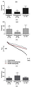
Comparison of SDH and MAO activities, MPTP opening, and decreases in A520 for the two groups. (a) Comparison of SDH activities in cardiac mitochondria from fasudil-treated, untreated diabetic, and control rats revealed statistically significant differences. SDH activities in cardiac mitochondria from fasudil-treated group were marked higher than that of untreated diabetic group (P = 0.003). (b) Comparison of MAO activities in cardiac mitochondria from fasudil-treated, untreated diabetic, and control rats revealed statistically significant differences. MAO activities in cardiac mitochondria from fasudil-treated group were significantly higher than that of untreated diabetic group (P = 0.015). (c) Effects of fasudil on MPTP opening (A520: absorbance at 520 nm). The trace represented the mean of A520 from three groups. The top trace represented cardiac mitochondria from SD control rats, the middle trace showed the absorbance in mitochondria from diabetic rats treated with fasudil, and lower traces showed the absorbance in mitochondria from untreated diabetic rats. (d) Comparison of decreases in A520 for the three groups. The decreases in A520 were significantly lower in diabetic rats treated with fasudil compared to untreated rats (P < 0.001).
Figure 3
Figure 3
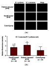
Effect of fasudil on transmembrane potential in mitochondria. Red fluorescence represents the mitochondrial aggregate form of JC-1 (JC-1 polymers), which indicates the intact mitochondrial membrane potential. Green fluorescence represents the monomeric form of JC-1 (JC-1 monomers), which indicates the dissipation of mitochondrial transmembrane potential. (a) Representative results of cardiac mitochondria stained with JC-1 and derived from fasudil-treated, untreated diabetic, and control rats. (b) Ratios of JC-1 monomer to JC-1 polymer (red/green fluorescence) for untreated diabetic rats, diabetic rats treated with fasudil, and control rats. Data are expressed as the mean ± SD. The ratio was significantly lower for diabetic rats treated with fasudil than for untreated diabetic rats (P = 0.037).
Figure 4
Figure 4
Effect of fasudil on cardiac Rho/ROCK activity. Levels of ROCK 1, ROCK 2, Rho A, and phosphorylated MYPT were detected in rat cardiac tissues using western blot analysis. (a) Representative results of assays of ROCK 1, ROCK 2, Rho A, and phospho-MYPT-1 and beta-actin abundances in rat cardiac tissues. (b) The levels of ROCK 1 protein expression were analyzed by western blot by using a polyclonal antibody to ROCK 1 to quantify its expressions in cardiac tissues. Data are expressed as the mean ± SD (P < 0.001 compared with untreated diabetic group). (c) The levels of ROCK 2 protein expression were analyzed by western blot by using a polyclonal antibody to ROCK 2 to quantify its expressions in cardiac tissues. Data are expressed as the mean ± SD (P < 0.001 compared with untreated diabetic group). (d) Average signal intensities quantified and expressed as percentage of the ratio of beta-actin to quantify Rho A expressions in cardiac tissues. Data are expressed as mean ± SD. The average signal intensities were significantly lower for diabetic rats treated with fasudil than for untreated diabetic rats (P = 0.008). (e) Average signal intensities quantified and expressed as a percentage of the ratio of T-MYPT to quantify levels of phosphorylated MYPT in cardiac tissues. Data are expressed as the mean ± SD. The ratio was significantly lower for diabetic rats treated with fasudil than for untreated diabetic rats (P = 0.019). (f) Five oxidative stress biomarkers were measured in heart tissues from diabetic-treated, untreated, and control rats. *P < 0.05 versus control group. # P < 0.05 versus untreated diabetic group.

References

    1. McBride HM, Neuspiel M, Wasiak S. Mitochondria: more than just a powerhouse. Current Biology. 2006;16(14):R551–R560. - PubMed
    1. Lesnefsky EJ, Moghaddas S, Tandler B, Kerner J, Hoppel CL. Mitochondrial dysfunction in cardiac disease: ischemia—reperfusion, aging, and heart failure. Journal of Molecular and Cellular Cardiology. 2001;33(6):1065–1089. - PubMed
    1. Walters AM, Porter GA, Jr., Brookes PS. Mitochondria as a drug target in ischemic heart disease and cardiomyopathy. Circulation Research. 2012;111(9):1222–1236. - PMC - PubMed
    1. Shen GX. Mitochondrial dysfunction, oxidative stress and diabetic cardiovascular disorders. Cardiovascular & Hematological Disorders—Drug Targets. 2012;12(2):106–112. - PubMed
    1. Shen GX. Oxidative stress and diabetic cardiovascular disorders: roles of mitochondria and NADPH oxidase. Canadian Journal of Physiology and Pharmacology. 2010;88(3):241–248. - PubMed

Publication types

MeSH terms

LinkOut - more resources