. 2013 Sep 1;443(2):197-207.
doi: 10.1016/j.virol.2013.05.014. Epub 2013 Jun 10.

Computational analysis of four human adenovirus type 4 genomes reveals molecular evolution through two interspecies recombination events

Affiliations

Computational analysis of four human adenovirus type 4 genomes reveals molecular evolution through two interspecies recombination events

Shoaleh Dehghan et al. Virology. .

Abstract

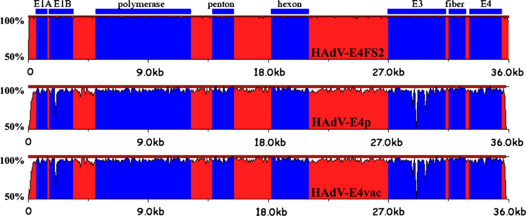
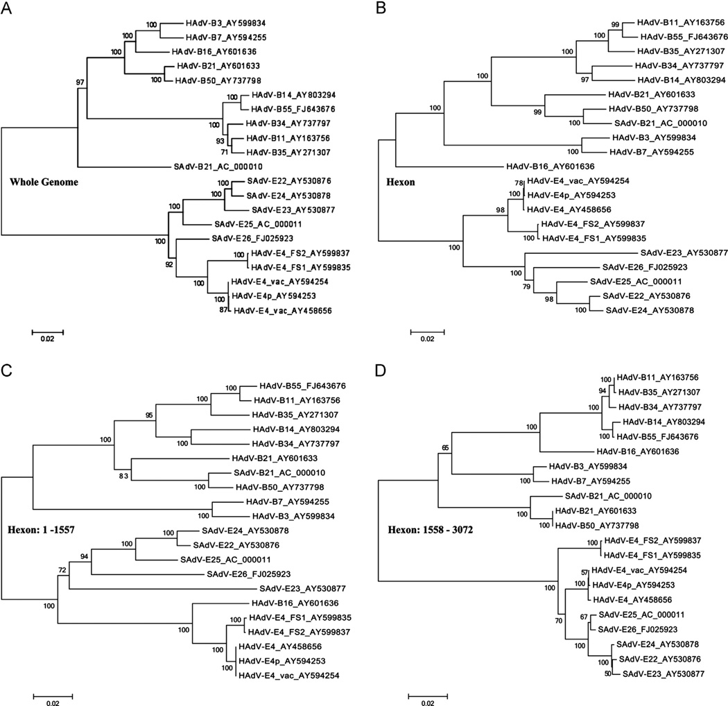
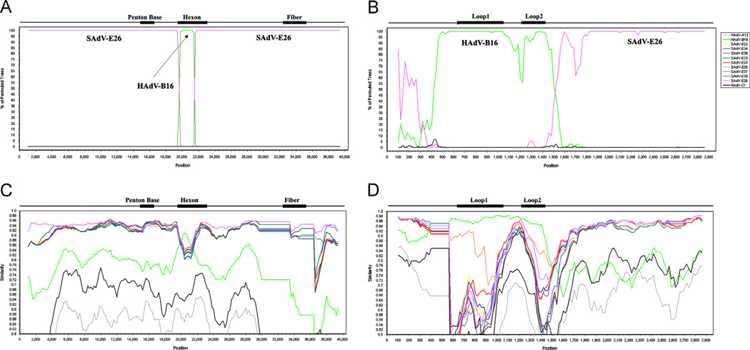
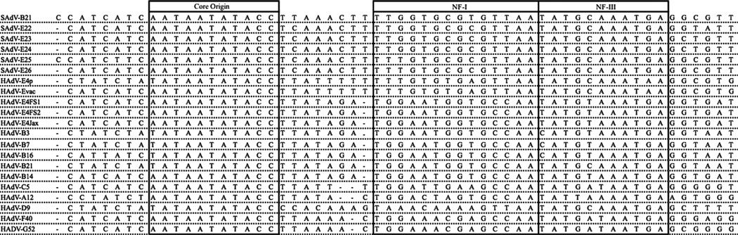
Computational analysis of human adenovirus type 4 (HAdV-E4), a pathogen that is the only HAdV member of species E, provides insights into its zoonotic origin and molecular adaptation. Its genome encodes a domain of the major capsid protein, hexon, from HAdV-B16 recombined into the genome chassis of a simian adenovirus. Genomes of two recent field strains provide a clue to its adaptation to the new host: recombination of a NF-I binding site motif, which is required for efficient viral replication, from another HAdV genome. This motif is absent in the chimpanzee adenoviruses and the HAdV-E4 prototype, but is conserved amongst other HAdVs. This is the first report of an interspecies recombination event for HAdVs, and the first documentation of a lateral partial gene transfer from a chimpanzee AdV. The potential for such recombination events are important when considering chimpanzee adenoviruses as candidate gene delivery vectors for human patients.

Keywords: Adenovirus; Molecular evolution; Recombination; Zoonosis.


Figures

Fig. 1
Fig. 1
Pairwise genome comparative analysis. zPicture (http://zpicture.dcode.org/) utilizes a local alignment algorithm, BlastZ, to display the regions of nucleotide sequence similarity between HAdV-E4FS1 and query genomes: HAdV-E4FS2 (top); HAdVE4p (middle); and HAdV-E4vac (bottom). Of note are the divergences at the ends of the genomes, corresponding to the semi-conserved inverted terminal repeat (ITR) sequences that contain critical functions, including DNA replication. The genome sequences are arrayed on the horizontal and the percent identity of genome pairs from 50% to 100% is noted along the y-axis. The colors are arbitrary and are used to provide contrast: blue regions highlight select genes or genome regions (E3), noted above the alignments. Red regions include both coding and noncoding sequences.
Fig. 2
Fig. 2
Phylogenomic analysis of HAdV genomes and hexons from species B and E, including select SAdVs. Bootstrap-confirmed neighbor-joining phylogenetic trees of the whole-genome (A) and hexon gene sequences are presented, with the hexon gene (B) further dissected for the detailed analyses of two domains: (C) nucleotides 1 to 1557, representing the proximal sequences and including the variable region and epitopic loopsL1 and L2, and (D) nucleotides 1558 to 3072, representing the remaining distal portion and including the conserved region. All phylogenetic trees were generated after multiple sequence alignments using the MAFFT software (http://www.ebi.ac.uk/tools/mafft) and default parameters. Bootstrapped, neighbor-joining trees with 1000 replicates were constructed using MEGA4.0.2 software with a maximum-composite-likelihood model. Bootstrap numbers shown at the nodes indicate the percentages of 1,000 replications producing the clade, with values above 80 considered robust. The scale bar is in units of nucleotide substitutions per site.
Fig. 3
Fig. 3
Nucleotide sequence recombination analysis of the whole genome and the hexon gene of HAdV-E4FSl. The genome of HAdV-E4FSl (AY599837) was analyzed for sequence recombination events with HAdV and SAdV genomes using the software Simplot, which is available on-line (http://sray.med.som.jhmi.edu/SCRoftware/simplot/). Bootscan analysis of the genome (A) and hexon (B) shows a lateral transfer of a portion of the hexon gene from HAdV-B 16. These are supported by the Simplot analysis of the genome (C) and hexon (D). Unlike the Simplot panels (C and D), only the SAdV-E26 genome, rather than the entire set of SAdV-E22 through -E26 sequences, is presented in the Bootscan panels (A and B), as similar genomes compete out the signal. For Bootscan, removing four of the five genomes and repeating the analysis with each individually, gives a clearer representation, reflecting the Simplot result for the highest similarity match. MAFFT software was used to align the sequences prior to recombination analysis (http://mafft.cbrc.jp/alignment/server/). Default parameter settings for the Simplot software were used for analyzing the hexon sequences: window size (200 nucleotides [nt]), step size (20 nt), replicates used (n=100), gap stripping (on), distance model (Kimura) and tree model (neighbor-joining). Similarly, whole genomes were analyzed, beginning with an initial alignment using MAFFT and following with recombination analysis using Simplot and Bootscan. Only the window size and step size were altered for the genome analysis (1000 and 200, respectively), with the rest of the default parameters unchanged. Genome nucleotide positions are noted along the x-axis, and the percentages of permutated trees that supported grouping are marked along the y-axis. For reference, select genome and gene-specific landmarks are noted above each graph. Colors: pink, SAdV-E26 and green, HAdV-B16.
Fig. 4
Fig. 4
Multiple sequence alignment of nucleotide sequences from the inverted terminal repeat (ITR). Edited Clustal Omega-generated (www.ebi.ac.uk/Tools/msa/clustalo/) sequence alignments of ca. 58 bases from the 5’-termini of five HAdV-E4 genomes are compared with representative counterparts from other HAdV species and several SAdVs. For reference, the three sequence motifs implicated in adenovirus DNA replication are labeled and boxed: (1) “core origin”; (2) NF-I transcription factor binding motif; and (3) NF-III transcription factor binding motif. The Clustal Omega-generated alignment was manually edited to position gaps to generate an optimal alignment, using the SAdV-B21 as reference.

References

    1. Adrian T, Wadell G, Hierholzer JC, Wigand R. DNA restriction analysis ol adenovirus prototypes 1 to 41. Arch. Virol. 1986;91:277–290. - PubMed
    1. Aoki K, Benko M, Davison AJ, Echavarria M, Erdman DD, Harrach B, Kajon AE, Schnurr D, Wadell G. Toward an integrated human adenovirus designation system that utilizes molecular and serological data and serves both clinical and fundamental virology. J. Virol. 2011;85:5703–5704. - PMC - PubMed
    1. Bailey A, Mautner V. Phylogenetic relationships among adenovirus serotypes. Virology. 1994;205:438–52. - PubMed
    1. Barrero PR, Valinotto LE, Tittarelli E, Mistchenko AS. Molecular typing of adenoviruses in pediatric respiratory infections in Buenos Aires, Argentina (1999–2010) J. Clin. Virol. 2012;53:145–150. - PubMed
    1. Basnight M, Jr., Rogers NG, Gibbs CJ, Jr., Gajdusek DC. Characterization of four new adenovirus serotypes isolated from chimpanzee tissue explants. Am. J. Epidemiol. 1971;94:166–171. - PubMed

Publication types

LinkOut - more resources