. 2013 Jul;19(7):1066-73.
doi: 10.3201/eid1907.121094.

Mutation in spike protein cleavage site and pathogenesis of feline coronavirus

Affiliations

Mutation in spike protein cleavage site and pathogenesis of feline coronavirus

Beth N Licitra et al. Emerg Infect Dis. 2013 Jul.

Abstract

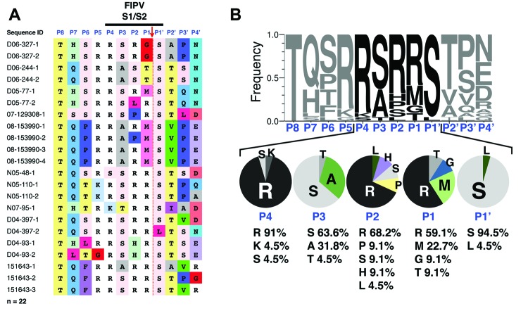
Feline coronaviruses (FCoV) exist as 2 biotypes: feline enteric coronavirus (FECV) and feline infectious peritonitis virus (FIPV). FECV causes subclinical infections; FIPV causes feline infectious peritonitis (FIP), a systemic and fatal disease. It is thought that mutations in FECV enable infection of macrophages, causing FIP. However, the molecular basis for this biotype switch is unknown. We examined a furin cleavage site in the region between receptor-binding (S1) and fusion (S2) domains of the spike of serotype 1 FCoV. FECV sequences were compared with FIPV sequences. All FECVs had a conserved furin cleavage motif. For FIPV, there was a correlation with the disease and >1 substitution in the S1/S2 motif. Fluorogenic peptide assays confirmed that the substitutions modulate furin cleavage. We document a functionally relevant S1/S2 mutation that arises when FIP develops in a cat. These insights into FIP pathogenesis may be useful in development of diagnostic, prevention, and treatment measures against coronaviruses.

Keywords: FECV; FIP; FIPV; Macrophage; conserved furin cleavage motif; feline coronavirus; feline enteric coronavirus; feline infectious peritonitis; feline infectious peritonitis virus; pathology; protease; protein processing; viruses.


Figures

Figure 1
Figure 1
Sequence analysis of feline enteric coronavirus (FECV) spike S1/S2 site. RNA from 30 FECVs collected from 30 fecal samples obtained from subclinically infected cats was extracted, purified, and reverse-transcribed into cDNA. Sequencing of the spike gene was performed in a region surrounding the S1/S2 cleavage site. A) Sequence alignment. Sequence identification row (blue font): residue positions in the S1/S2 cleavage site from P8 to P4′. Red arrow indicates the site of furin cleavage. B) To visualize the diversity of residues at each position of the S1/S2 site, sequences were subjected to WebLogo 3.1 analysis (http://weblogo.threeplusone.com/create.cgi). Top: WebLogo for the 30 FECV S1/S2 sequences with the frequency of residue found at each position displayed. Bottom: summary of the diversity of residues for each position from P4 to P1′ and percentages of each amino acid represented.
Figure 2
Figure 2
Sequence analysis of feline infectious peritonitis virus (FIPV) spike S1/S2 site. RNA from 22 FIPVs collected from 11 cats who had feline infectious peritonitis was extracted, purified, and reverse-transcribed into cDNA. Sequencing of the spike gene was performed in a region surrounding the S1/S2 cleavage site. A) Sequence alignment. Sequence identification row (blue font): residue positions in the S1/S2 cleavage site from P8 to P4′. Red arrow indicates the site of furin cleavage. B) To visualize the diversity of residues at each position of the S1/S2 site, sequences were subjected to WebLogo 3.1 analysis (http://weblogo.threeplusone.com/create.cgi). Top: WebLogo for the 22 FIPV S1/S2 sequences with the frequency of residue found at each position displayed. Bottom: summary of the diversity of residues for each position from P4 to P1′ and percentages of each amino acid represented.
Figure 3
Figure 3
Amino acid substitution frequency at each position of the feline infectious peritonitis virus S1/S2 cleavage site. The histogram is based on feline infectious peritonitis virus S1/S2 WebLogo 3.1 analysis (http://weblogo.threeplusone.com/create.cgi), showing percentage of modification of residues at each position of the S1/S2 site, compared with feline enteric coronavirus S1/S2 canonical sequence consensus.
Figure 4
Figure 4
Furin cleavage assays of fluorogenic peptides. A) Synthetic fluorogenic peptides were generated with sequences matching consensus feline enteric coronavirus and a panel of modified sequences with substitutions (shown in red) found by feline infectious peritonitis virus sequencing. Peptides (50 μmol/L) were subjected to cleavage by recombinant human furin (1 U/100 μL), at pH 7.5, 30°C, and the release of fluorescence over time was measured by a spectrofluorometer enabling calculation of the Vmax of each reaction. Peptide cleavage scores generated by the ProP 1.0 server (www.cbs.dtu.dk/services/ProP/) are also displayed. B) For each modified peptide (substitutions shown in red), the percentage of cleavage rate compared with the canonical sequence was calculated and displayed. Cleavage assays were performed in >3 independent experiments. Error bars indicate SD for each measurement.

References

    1. Haijema BJ, Rottier PJ, de Groot RJ. Feline coronaviruses: a tale of two-faced types. In: Thiel V, editor. Coronaviruses: molecular and cellular biology. Norfolk (UK): Caister Academic Press; 2007. p. 183–203.
    1. King AMQ, Lefkowitz E, Adams MJ, Carstens EB. Virus taxonomy: IXth Report of the International Committee on Taxonomy of Viruses. London:Elsevier; 2011.
    1. Hohdatsu T, Okada S, Ishizuka Y, Yamada H, Koyama H. The prevalence of types I and II feline coronavirus infections in cats. J Vet Med Sci. 1992;54:557–62 . 10.1292/jvms.54.557 - DOI - PubMed
    1. Benetka V, Kubber-Heiss A, Kolodziejek J, Nowotny N, Hofmann-Parisot M, Mostl K. Prevalence of feline coronavirus types I and II in cats with histopathologically verified feline infectious peritonitis. Vet Microbiol. 2004;99:31–42 . 10.1016/j.vetmic.2003.07.010 - DOI - PMC - PubMed
    1. Shiba N, Maeda K, Kato H, Mochizuki M, Iwata H. Differentiation of feline coronavirus type I and II infections by virus neutralization test. Vet Microbiol. 2007;124:348–52. 10.1016/j.vetmic.2007.04.031 - DOI - PMC - PubMed

Publication types

Substances

LinkOut - more resources