. 2013 Jun 10;23(6):826-38.
doi: 10.1016/j.ccr.2013.05.002.

SYK inhibition modulates distinct PI3K/AKT- dependent survival pathways and cholesterol biosynthesis in diffuse large B cell lymphomas

Affiliations

SYK inhibition modulates distinct PI3K/AKT- dependent survival pathways and cholesterol biosynthesis in diffuse large B cell lymphomas

Linfeng Chen et al. Cancer Cell. .

Abstract

B cell receptor (BCR) signaling pathway components represent promising treatment targets in diffuse large B cell lymphoma (DLBCL) and additional B cell tumors. BCR signaling activates spleen tyrosine kinase (SYK) and downstream pathways including PI3K/AKT and NF-κB. In previous studies, chemical SYK blockade selectively decreased BCR signaling and induced apoptosis of BCR-dependent DLBCLs. Herein, we characterize distinct SYK/PI3K-dependent survival pathways in DLBCLs with high or low baseline NF-κB activity including selective repression of the pro-apoptotic HRK protein in NF-κB-low tumors. We also define SYK/PI3K-dependent cholesterol biosynthesis as a feed-forward mechanism of maintaining the integrity of BCRs in lipid rafts in DLBCLs with low or high NF-κB. In addition, SYK amplification and PTEN deletion are identified as selective genetic alterations in primary "BCR"-type DLBCLs.


Figures

Figure 1
Figure 1. Cellular proliferation of DLBCL cell lines following SYK depletion
(A) The SYK protein level in cell lysates prepared from the indicated cell lines transduced with a negative control (NC) shRNA or the indicated SYK shRNAs was assessed by immunoblotting. β-actin, loading control. (B) Cellular proliferation of SYK-depleted DLBCL cell lines was measured 72 hr after puromycin selection by MTS assay. The proliferation of SYK-depleted cells is relative to that of NC cells. Error bars represent standard deviation (SD) of three independent assays from a representative experiment. p-values for NC vs. each SYK shRNA were determined with a one-sided Welch t-test. ***, p≤0.0001; **, p≤0.001. See also Figure S1.
Figure 2
Figure 2. SYK and PI3K-dependent BCR signaling in DLBCLs with high baseline NF-κB
(A) NF-κB (p65) activity was determined in vehicle and R406-treated DLBCL cell lines. Pos. Ctrl.: Manufacturer’s positive control. BCR-dependent NF-κB-low lines, purple; BCR-dependent NF-κB-high lines, blue; BCR-independent OxPhos lines, green. (B) GSEA of NF-κB targets was performed in vehicle- vs. R406-treated DLBCL cell lines with high baseline NF-κB activity (HBL-1 and U-2932). The 19K genes in the genome were sorted from highest (left, red) to lowest (right, blue) relative expression in vehicle- vs. R406-treated lines (horizontal axis). Note that the positions of the NF-κB targets (Set #1, (Davis et al., 2010) Set #2, (Schreiber et al., 2006)) were significantly skewed toward the right end of the sorted list, reflecting their statistically significant downregulation in R406-treated lines. (C) Relative abundance of NF-κB target genes in vehicle- vs. R406-treated DLBCL cell lines with high baseline NF-κB activity was displayed with a colometric scale. NF-κB target genes derived from the GSEA leading edge, Set #1 (Davis et al., 2010). (D) BCL2A1 transcript abundance in DLBCL cell lines treated with vehicle or R406 (24 hr) was determined by qRT-PCR relative to PPIA. (E) BCL2A1 protein abundance in vehicle- and R406-treated DLBCL cell lines was assessed by immunoblotting. β-actin, loading control. (F) BCL2A1 transcript abundance in SYK-depleted DLBCL cell lines (72 hr following completion puromycin selection) was determined by qRT-PCR relative to PPIA. (G) NF-κB (p65) activity was determined in DLBCL cell lines treated with the PI3K inhibitor LY294002 or vehicle. (H) BCL2A1 transcript abundance in DLBCL cell lines treated with LY294002, R406, or vehicle (24 hr) was assessed by qRT-PCR relative to PPIA. In (A), (D), (F), (G) and (H), p-values for control vs. treated were determined with a one-sided Welch t-test. ***, p <0.0001; **, p <0.001; *, p <0.01. Error bars represent the SD of 3 independent assays in a representative experiment. See also Figure S2 and Table S1.
Figure 3
Figure 3. SYK and PI3K-dependent signaling in DLBCLs with low baseline NF-κB
(A, B) DHL4, DHL6 and LY7 cell lines were retrovirally transduced with mAKT or pMIG vector, FACS-sorted for GFP expression, treated for 24 hr with R406 or vehicle, then analyzed for p-AKT(S473) and total AKT by immunoblotting (A) and for proliferation (B). (C) DLBCL cell lines were incubated with R406 or vehicle for 1 hr, BCR cross-linked (BCR) or left untreated, fixed, then stained with DAPI (blue) and with Cy3-conjugated anti-FOXO1 antibody (red) (left panels). Scale bar represents 10 μm. The mean nuclear staining intensity of Cy3-conjugated-FOXO1 was quantified for each cell and plotted against its mean DAPI staining intensity (right panels). (D) The transcript abundance of FOXO1 target genes, p27 and BIM, in DLBCL cell lines treated with R406 or vehicle for 24 hr was assessed by qRT-PCR relative to PPIA. p-values for mAKT vs. pMIG (control) at each timepoint (B) and for vehicle vs. R406 (D) were determined with a one-sided Welch t-test. ***, p≤0.0001; **, p≤0.001. Error bars represent the SD of 3 independent assays in a representative experiment. See also Figure S3.
Figure 4
Figure 4. HRK induction in DLBCL cell lines following SYK or PI3K inhibition
(A) HRK transcript abundance in DLBCL cell lines following R406 treatment (24 hr) was determined by qRT-PCR. (B) HRK expression in SYK-depleted DLBCL cell lines was assessed by qRT-PCR. (C) HRK expression in the indicated cell lines following treatment with R406, LY294002, or vehicle for 24 hr assessed by qRT-PCR. (D) HRK transcript abundance in DLBCL lines transduced with mAKT or pMIG, FACS-sorted, then treated with R406 or vehicle for 24 hr was analyzed by qRT-PCR. (E) DHL4 cells stably transduced with the indicated HRK siRNA or negative control siRNA (NC) were treated with R406 or vehicle for 24 hr and analyzed for HRK transcript abundance by qRT-PCR. (F) Apoptosis of DHL4 cells transduced with HRK siRNA or negative control siRNA (NC) and treated with vehicle or R406 for 72 hr was assessed by Annexin V/PI staining. (G and H) Viable DLBCL tumor cells isolated from cryopreserved primary patient samples were analyzed for cell surface Ig expression (G) or for HRK and BCL2A1 transcript abundance after treatment with R406 or vehicle for 24 hr by qRT-PCR (H). p-values for vehicle vs. R406 (A and H), vehicle vs. R406 or LY29004 (C), NC vs. shSYK (B) and pMIG +R406 vs. mAKT +R406 (D) were determined with a one-sided Welch t-test. ***, p≤0.0001; **, p≤0.001; *, p≤0.01. Error bars represent the SD of 3 independent assays in a representative experiment. See also Figure S4.
Figure 5
Figure 5. SYK expression following chemical SYK inhibition and FOXO1 depletion
(A) SYK transcript abundance in DLBCL cell lines was assessed by qRT-PCR following 24 hr of treatment with R406 or vehicle. (B) SYK transcript abundance in DLBCL cell lines treated with R406 or vehicle for 24 hr following transduction with indicated shRNA was analyzed by qRT-PCR. p-values for vehicle vs. R406 were determined with a one-sided Welch t-test. ***, p≤0.0001; **, p≤0.001; *, p≤0.01, #, p≤0.05. Error bars represent the SD of 3 independent assays in a representative experiment. See also Figure S5.
Figure 6
Figure 6. Cholesterol biosynthesis in DLBCLs following SYK or PI3K inhibition
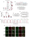
(A) Components of the cholesterol biosynthesis pathway modulated by R406 treatment (red) were identified by pathway analyses of the profiled cell lines. (B) HMGCS1 protein abundance in R406- or vehicle-treated DLBCL cell lines was assessed by immunoblotting. β-actin, loading control. (C) HMGCS1 transcript abundance in DLBCL cell lines transduced with indicated shRNA was assessed by qRT-PCR. p-values for NC vs. shSYK were determined with a one-sided Welch t-test. ***, p≤0.0001; *, p≤0.01; #, p≤0.02. Error bars represent the SD of 3 independent assays in a representative experiment. (D) HMGCS1 protein abundance in DHL4 following treatment with R406 or LY294002 was assessed by immunoblotting. β-actin, loading control. (E) HMGCS1 and p-AKT(S473) in parental DHL4 cells and DHL4 cells transduced with mAKT or pMIG and treated with R406 was determined by immunoblotting. β-actin, loading control. (F) Membrane cholesterol content in DLBCL cell lines treated with vehicle (black) or R406 (red) for 2 days and fixed with 4% paraformaldehyde was assessed by filipin flow cytometry. Unstained controls in gray. (G) DHL4 and LY7 cells were incubated with vehicle or R406 for 48 hr and left untreated (-BCR) or stimulated with anti-Ig for 10 min (+BCR). The cells were fixed, stained with filipin (green) and the indicated Cy3-conjugated anti-Ig (red) and visualized by confocal microscopy. Scale bar represents 25 μm. See also Figure S6.
Figure 7
Figure 7. SYK and PTEN copy number alterations in primary DLBCLs
(A) SYK and PTEN copy numbers were assessed and associated with SYK and PTEN transcript levels in primary DLBCLs (n = 169). SYK and PTEN copy numbers and transcript abundance were derived from (Monti et al., 2012). p-values were determined with a one-sided t-test. Box plot (median, line; 25% and 75% quartile, box; whiskers, maximum to minimum). (B) Relative frequency of SYKAMP and PTENDEL was assessed in transcriptionally defined “BCR” type vs. other primary DLBCLs (n = 147 [tumors with high confidence classification into CCC categories]) (Monti et al., 2012). p-values were determined with two-sided Fisher exact test. See also Figure S7 and Table S2.
Figure 8
Figure 8. SYK and PI3K-dependent viability pathways in DLBCLs
(A) SYK- and PI3K-dependent anti-apoptotic pathways in BCR-dependent DLBCLs. In BCR-dependent DLBCLs with low baseline NF-κB activity, SYK or PI3K inhibition relieve BCR-dependent repression of the pro-apoptotic BH3 molecule, HRK. In BCR-dependent DLBCLs with high baseline NF-κB activity, SYK or PI3K inhibition decrease the abundance of the anti-apoptotic NF-κB targets such as BCL2A1. (B) SYK- and PI3K-dependent regulation of cholesterol biosynthesis and associated integrity of BCRs in lipid rafts in BCR-dependent DLBCLs.

References

    1. Bengoechea-Alonso M, Ericsson J. SREBP in signal transduction: cholesterol metabolism and beyond. Current Opinion in Cell Biology. 2007;19:215–222. - PubMed
    1. Caro P, Kishan AU, Norberg E, Chapuy B, Stanley I, Tondera D, Gounarides J, Zhang B, Green M, Chen L, et al. Metabolic signatures uncover novel targets in molecular subsets of diffuse large B cell lymphoma. Cancer Cell. 2012;22:547–560. - PMC - PubMed
    1. Chen L, Juszczynski P, Takeyama K, Aguiar RC, Shipp MA. Protein tyrosine phosphatase receptor-type O truncated (PTPROt) regulates SYK phosphorylation, proximal B-cell-receptor signaling, and cellular proliferation. Blood. 2006;108:3428–3433. - PubMed
    1. Chen L, Monti S, Juszczynski P, Chen W, Kutok JL, Shipp MA. SYK-dependent tonic B-cell receptor signaling is a rational treatment target in diffuse large B-cell lymphoma. Blood. 2008;111:2230–2237. - PMC - PubMed
    1. Cheng S, Coffey G, Zhang XH, Shaknovich R, Song Z, Lo P, Pandey A, Melnick AM, Somma U, Wang L. SYK inhibition and response prediction in diffuse large B-cell lymphoma. Blood. 2011;118:6342–6352. - PubMed

Publication types

MeSH terms

Associated data