. 2013 Jul 12;436(4):660-5.
doi: 10.1016/j.bbrc.2013.06.012. Epub 2013 Jun 11.

Piperlongumine inhibits LMP1/MYC-dependent mouse B-lymphoma cells

Affiliations

Piperlongumine inhibits LMP1/MYC-dependent mouse B-lymphoma cells

Seong-Su Han et al. Biochem Biophys Res Commun. .

Abstract

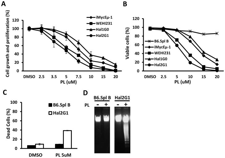
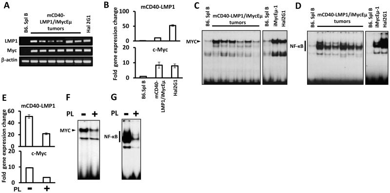
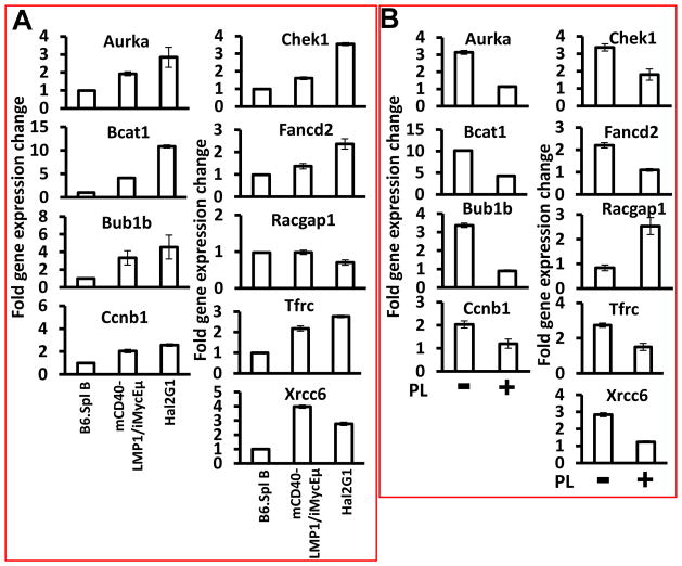
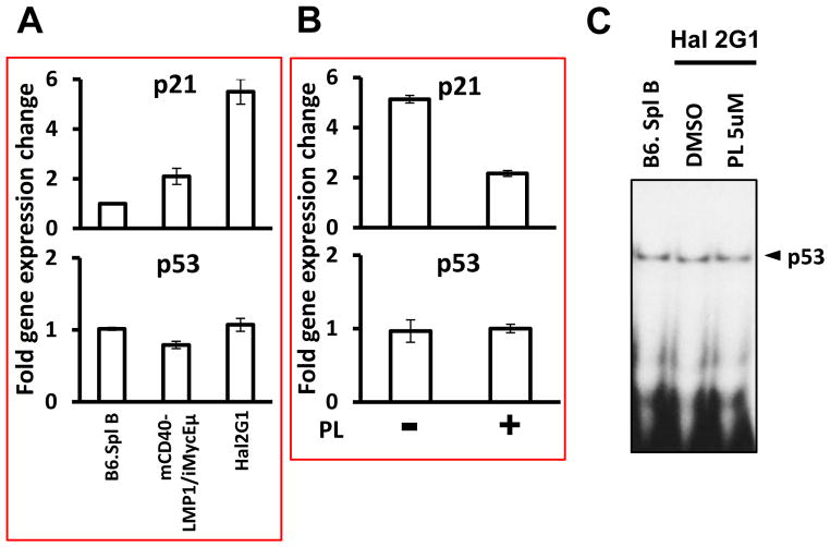
Piperlongumine (PL), isolated from the fruit of Long pepper, Piper longum, is a cancer-inhibiting compound that selectively kills tumor cells while sparing their normal counterparts. Here we evaluated the efficacy with which PL suppresses malignant B cells derived from a newly developed, double-transgenic mouse model of human endemic Burkitt lymphoma (BL), designated mCD40-LMP1/iMyc(Eμ). PL inhibited tumor cell proliferation in a concentration-dependent manner and induced apoptosis of neoplastic but not normal B cells. Treatment with PL resulted in downregulation of EBV-encoded LMP1, cellular Myc, constitutive NF-κB activity, and a host of LMP1-Myc-NF-κB-regulated target genes including Aurka, Bcat1, Bub1b, Ccnb1, Chek1, Fancd2, Tfrc and Xrcc6. Of note, p21(Cip1)-encoding Cdkn1a was suppressed independent of changes in Trp53 mRNA levels and p53 DNA-binding activity. Considering the central role of the LMP1-NF-κB-Myc axis in B-lineage neoplasia, these findings further our understanding of the mechanisms by which PL inhibits B-lymphoma and provide a preclinical rationale for the inclusion of PL in new interventions in blood cancers.

Keywords: BL; Burkitt lymphoma; Cancer therapy and prevention; Epstein Barr virus; NF-κB; PL; Piperlongumine; Transgenic mouse model of human endemic Burkitt lymphoma; p21-Encoding Cdkn1a.


Figures

Figure 1
Figure 1. PL-dependent growth inhibition and apoptosis
(A) MTS assay, a measure of metabolic activity, was used as surrogate for cell proliferation. Cells (106/ml) were treated with the indicated concentrations of PL for 24 hrs. Results were normalized to cells treated with DMSO only (solvent control). Error bars indicate standard deviations of the mean determined in a representative experiment performed in triplicate. IC50 of PL was determined for each cell line and used for all following experiments unless otherwise stated. (B) Cells were treated with PL as described in panel A and the fraction of dead cells was determined with the assistance of the TBE assay. (C) Normal B splenocytes (black bars) or Hal2G1 cells (white bars) were treated for 24 hrs with 5 μM PL or left untreated (DMSO only). Cells were stained with PI (propodium iodide) and subjected to flow cytometry to determine the fraction of dead cells containing sub-G1 DNA content. Means and standard deviations (error bars) were determined in three independent experiments and plotted. Asterisk indicates statistical significance at p < 0.05 using the t test. (D) DNA laddering of genomic DNA was readily apparent in PL-treated (+) Hal2G1 cells but not in Hal2G1 cells left untreated (−) or normal (treated or untreated) splenic B cells.
Figure 2
Figure 2. PL inhibits the LMP1- NF-κB-Myc pathway
(A) RT-PCR analysis of LMP1 and Myc expression in normal splenic B cells (lane 1), B-cell tumors obtained from 6 different mCD40-LMP1/iMyc-transgenic mice (lanes 2–7) and Hal2G1 cells (lane 8). The housekeeping gene, Actb, was used as control. (B) qPCR analysis of the samples from panel A. PCR results were normalized to Hprt message levels and converted to fold gene expression change by dividing the normalized value from malignant B cells to that of normal B cells. (C) EMSA of nuclear extracts prepared from normal B cells, lymphoma tissues and cell lines as indicated. Myc DNA-binding activity is indicated by labeled arrowhead. (D) EMSA of the samples from panel C. NF-κB DNA-binding activity is indicated by thick vertical line. (E) PL-dependent reduction of LMP1 and Myc mRNA levels in Hal2G1 cells, as determined by qPCR. Cells were cultured in presence (+) or absence (−) of PL for 24 hrs. (F) NF-κB DNA-binding activity in Hal2G1 cells treated with PL as shown in panel E (+) or left untreated (−). (G) Myc DNA-binding activity in Hal2G1 cells treated with PL as shown in panel E (+) or left untreated (−).
Figure 3
Figure 3. PL-dependent expression changes of LMP1-NF-κB-Myc target genes
(A) qPCR results indicating elevated expression of all indicated genes except Racgap1 in lymphomas from mCD40-LMP1/iMyc mice (n = 6) or Hal2G1 cells relative to normal B220+ splenocytes. Target gene expression was normalized to Hprt and converted to fold gene expression change by dividing the results from lymphomas or Hal2G1 cells to the value of normal B cells. Error bars represent the standard deviation of the mean from triplicate measurements. (B) qPCR results showing that compared to Hal2G1 cells left untreated (−), all indicated genes except Racgap1 were down regulated upon treatment with PL for 24 hrs (+). Racgap1 was up regulated.
Figure 4
Figure 4. Significant PL-dependent downregulation of p21-encoding Cdkn1a message in the face of unchanged p53 DNA-binding activity
(A) qPCR results indicating elevated expression of Cdkn1a in mCD40-LMP1/iMyc-induced lymphomas (n = 6) or Hal2G1 cells compared to normal B220+ splenocytes. Cdkn1a expression was normalized to Hprt and converted to fold gene expression change by dividing the values from lymphomas or Hal2G1 cells to the value of normal B cells. Error bars represent the standard deviation of the mean from triplicate measurements. (B) qPCR results showing that compared to Hal2G1 cells left untreated (−), Cdkn1a was down regulated in cells treated with PL for 24 hrs (+). (C) EMSA showing no difference in DNA binding activity of p53 in normal B cells (lane 1) and Hal2G1 cells cultured in absence (lane 2) or presence of 5 μM PL for 24 hrs.

References

    1. Singh N, Kumar S, Singh P, Raj HG, Prasad AK, Parmar VS, Ghosh B. Piper longum Linn. Extract inhibits TNF-α-induced expression of cell adhesion molecules by inhibiting NF-κB activation and microsomal lipid peroxidation. Phytomedicine. 2008;15:284–291. - PubMed
    1. Han SS, Son DJ, Yun H, Kamberos NL, Janz S. Piperlongumine inhibits proliferation and survival of Burkitt lymphoma in vitro. Leukemia Research. 2013;37:146–154. - PMC - PubMed
    1. Raj L, Ide T, Gurkar AU, Foley M, Schenone M, Li X, Tolliday NJ, Golub TR, Carr SA, Shamji AF, Stern AM, Mandinova A, Schreiber SL, Lee SW. Selective killing of cancer cells by a small molecule targeting the stress response to ROS. Nature. 2011;475:231–234. - PMC - PubMed
    1. Wang D, Liebowitz D, Kieff E. An EBV membrane protein expressed in immortalized lymphocytes transforms established rodent cells. Cell. 1985;43:831–840. - PubMed
    1. Dalla-Favera R, Bregni M, Erikson J, Patterson D, Gallo RC, Croce CM. Human c-myc onc gene is located on the region of chromosome 8 that is translocated in Burkitt lymphoma cells. Proceedings of the National Academy of Sciences. 1982;79:7824–7827. - PMC - PubMed

Publication types