. 2013 Aug 15;73(16):5232-41.
doi: 10.1158/0008-5472.CAN-13-0470. Epub 2013 Jun 13.

Manganoporphyrins increase ascorbate-induced cytotoxicity by enhancing H2O2 generation

Affiliations

Manganoporphyrins increase ascorbate-induced cytotoxicity by enhancing H2O2 generation

Malvika Rawal et al. Cancer Res. .

Abstract

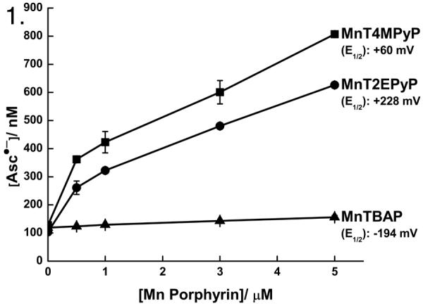
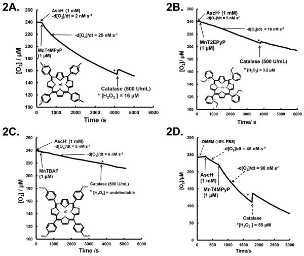
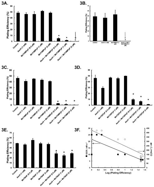
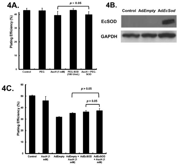
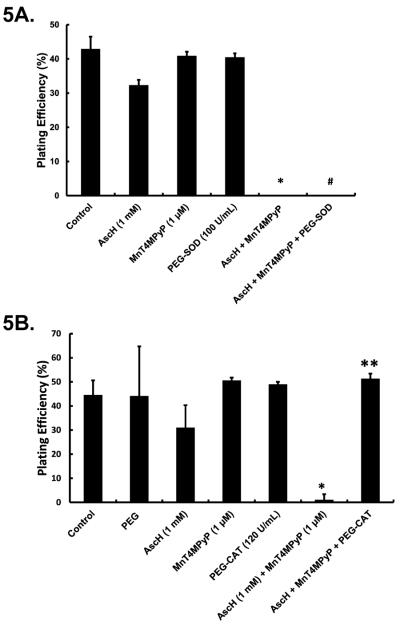
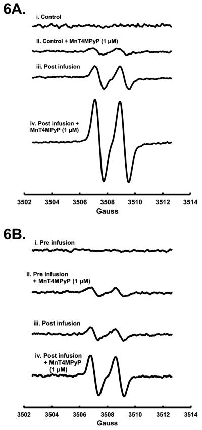
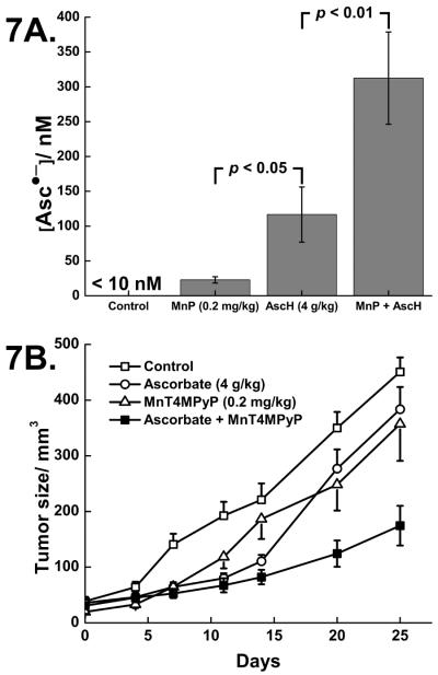
Renewed interest in using pharmacological ascorbate (AscH-) to treat cancer has prompted interest in leveraging its cytotoxic mechanism of action. A central feature of AscH- action in cancer cells is its ability to act as an electron donor to O2 for generating H2O2. We hypothesized that catalytic manganoporphyrins (MnP) would increase AscH- oxidation rates, thereby increasing H2O2 fluxes and cytotoxicity. Three different MnPs were tested (MnTBAP, MnT2EPyP, and MnT4MPyP), exhibiting a range of physicochemical and thermodynamic properties. Of the MnPs tested, MnT4MPyP exerted the greatest effect on increasing the rate of AscH- oxidation as determined by the concentration of ascorbate radical [Asc•-] and the rate of oxygen consumption. At concentrations that had minimal effects alone, combining MnPs and AscH- synergized to decrease clonogenic survival in human pancreatic cancer cells. This cytotoxic effect was reversed by catalase, but not superoxide dismutase, consistent with a mechanism mediated by H2O2. MnPs increased steady-state concentrations of Asc•- upon ex vivo addition to whole blood obtained either from mice infused with AscH- or patients treated with pharmacologic AscH-. Finally, tumor growth in vivo was inhibited more effectively by combining MnT4MPyP with AscH-. We concluded that MnPs increase the rate of oxidation of AscH- to leverage H2O2 flux and ascorbate-induced cytotoxicity.


Figures

Figure 1
Figure 1. Manganoporphyrins increase ascorbate radical indicating an increase in the rate of AscH oxidation
As seen by EPR spectroscopy, addition of MnPs to a solution of AscH (1 mM) in PBS, increased [Asc•−] in a dose-dependent manner. This increase is dependent on the reduction potential of the individual MnPs. At lower concentrations of Asc•− the curves have a greater slope that decreases as [Asc•−] increases, which is consistent with an increased rate of AscH oxidation and the second-order nature of the dismutation reaction of Asc•− [14]. n = 3.
Figure 2
Figure 2. Manganoporphyrins increase the rate of oxygen consumption, leading to generation of H2O2
A. AscH consumes oxygen at the rate of 2–5 nM s−1 in PBS. Addition of MnT4MPyP (1.0 μM) increases the rate of oxygen consumption to 22–25 nM s−1. Addition of catalase (500 U/mL) leads to a return of O2 with 16–20 μM H2O2 accumulating in solution. n = 3. Inset: MnT4MPyP molecular structure. B. Addition of MnT2EPyP (1.0 μM) to AscH (1 mM) solution increases the oxygen consumption rate from ≈ 5 nM s−1 to ≈ 10 nM s−1. Addition of catalase indicates 3–5 μM of H2O2 has accumulated in solution after 60 min. n = 3. Inset: MnT2EPyP molecular structure. C. MnTBAP (1.0 μM) addition to AscH (1.0 mM) solution does not alter the oxygen consumption rate. Addition of catalase does not return detectable amounts of oxygen demonstrating minimal H2O2 accumulation. n = 3. Inset: MnTBAP molecular structure. D. In DMEM with 10 % FBS, oxygen consumption for AscH (1 mM) is 20–45 nM s−1. Addition of MnT4MPyP increases oxygen consumption to 80–100 nM s−1. Addition of catalase 30 min after MnT4MPyP leads to a return of oxygen, indicating that 50–60 μM H2O2 has accumulated in solution. n = 3.
Figure 3
Figure 3. Manganoporphyrins enhance AscH-induced cytotoxicity in pancreatic cancer cell lines
A. Treatment with AscH or MnT4MPyP alone does not alter clonogenic survival of MIA PaCa-2 cells. However, the combination of AscH (1 mM) and MnT4MPyP (0.5 μM) decreases plating efficiency to 5 ± 1 %. When MnT4MPyP (2 μM) was added to AscH, no clones survived. n = 3. *p < 0.001 vs. controls. B. MnT4MPyP (1 μM) or AscH (1 mM) did not affect AsPC-1 clonogenic survival while the combination led to no surviving clones. n = 3. *p < 0.001 vs. control. C. MnT4MPyP (0.5 – 2 μM) or AscH (1 mM) did not affect Panc-1 clonogenic survival. However, addition of MnT4MPyP (0.5 μM) to AscH (1 mM) decreased survival to 3 ± 0.2 %. Only 1 ± 0.1% clones survived exposure to 2 μM MnT4MPyP in the presence of AscH. n = 3. *p < 0.001 vs. control. D. MnT2EPyP (0.5, 1 or 2 μM) or AscH (1 mM) does not alter MIA PaCa-2 survival. MnT2EPyP (0.5 μM) combined with AscH (1 mM) decreases plating efficiency to 10 ± 0.5 %. When increased concentrations of MnT2EPyP are used, plating efficiency was reduced to ≤ 5 %. n = 3. *p < 0.001 vs. controls. E. MnTBAP (0.5, 1 or 2 μM) does not alter MIA PaCa-2 plating efficiency. However, when MnTBAP was combined with AscH, MIA PaCa-2 plating efficiency was decreased to 30 ± %. n = 3. *p < 0.001 vs. control. F. The OCR of AscH when combined with MnPs correlates with log (plating efficiency) in pancreatic cancer cell lines (solid line, R2 = 0.9, p < 0.05). In solution, [Asc•−] in the presence of MnPs (from Figure 1) strongly correlated with log (plating efficiency) in MIA PaCa-2, AsPC-1 and Panc-1 cells (hatched line, R2≈ 0.9, p < 0.05. The (2) below specific points on the graph indicate that two identical points at that value.
Figure 4
Figure 4. SOD does not alter AscH-induced cytotoxicity
A. Cells were treated with AscH (1 mM) and PEG-SOD (100 U/mL) alone or in combination for 60 min, washed and seeded for clonogenic survival. PEG-SOD did not alter clonogenic survival and combining PEG-SOD with AscH did not alter clonogenic survival of MIA PaCa-2 cells. p > 0.05 comparing AscHvs. AscH + PEG-SOD. B. MIA PaCa-2 cells transfected with AdEmpty or AdEcSOD were lysed and resolved on SDS-polyacrylamide gel and then blotted for the presence of EcSOD. GAPDH was used as a loading control. Lanes 1, 2 and 3 represent parental cells, cells infected with the AdEmpty vector and cells infected with the AdEcSOD vector, respectively. C. MIA PaCa-2 cells were transfected with either AdEmpty or AdEcSOD at 100 MOI. There were minimal changes (p > 0.05) in plating efficiency in AdEmpty or AdEcSOD infected cells treated with AscH.
Figure 5
Figure 5. H2O2 mediates MnP enhanced AscH-induced cytotoxicity
A. PEG-SOD (100 U/mL) did not reverse MnT4MPyP enhanced AscH-induced cytotoxicity in MIA PaCa-2 cells, suggesting that superoxide is not directly responsible for the observed cytotoxicity. n =3. *p < 0.001 vs. controls. #p > 0.05 vs. AscH + MnP. B. Treatment with PEG-CAT (120 U/mL) reverses MnT4MPyP enhanced AscH-induced cytotoxicity suggesting that H2O2 mediates the observed cytotoxicity. n = 3. *p < 0.001 vs. control. **p < 0.001 vs. AscH + MnT4MPyP treated cells.
Figure 6
Figure 6. MnT4MPyP enhances ascorbate radical concentration in whole blood as seen by EPR spectroscopy
A. In whole blood from mice, Asc•− is below the limit of detection. MnT4MPyP (1.0 μM) increased [Asc•−]ss to 97 nM. When mice were treated with AscH (4 g/kg) resulting in a plasma level of [AscH] of 29 mM, [Asc•−]ss was increased to 350 nM. With addition of MnT4MPyP (1.0 μM) to ascorbate treated mice, [Asc•−]ss increased more than 3-fold to 1200 nM. n = 3. (Hyperfine splitting of Asc•−, aH = 1.76 G.) B. In whole blood from humans, Asc•− is also below the limit of detection. [Asc•−]ss increases to 82 nM upon addition of MnT4MPyP (1.0 μM) to human whole blood. After infusion of pharmacological AscH(100 g), [AscH] increased to 22 mM and [Asc•−]ss increased to 120 nM. Upon addition of MnT4MPyP (1.0 μM), [Asc•−]ss increased to 360 nM. n = 3.
Figure 7
Figure 7. MnT4MPyP enhances ascorbate-induced cytotoxicity and ascorbate radical concentration in vivo
A. In whole blood from a separate group of mice, Asc•− is below the limit of detection. [Asc]ss increases to 22 nM upon treatment with MnT4MPyP (0.2 mg/kg i.p.). After i.p. injection of pharmacological AscH (4 g/kg), [AscH] increased to 30 mM and [Asc•−]ss increased to 120 nM. Combining both treatments increased [Asc•−]ss to 310 nM. n = 3 for each determination. AscHvs.MnT4MPyP, p < 0.05. AscHvs. AscH + MnT4MPyP, p < 0.01. B. MnT4MPyP combined with pharmacological ascorbate decreased MIA PaCa-2 tumor growth in nude mice. The MnT4MPyP and ascorbate-treated animals had significantly slower tumor growth when compared to the control and ascorbate groups (*#p < 0.05, n = 12–16/group, means ± SEM). MIA PaCa-2 tumor cells (2 × 10⁶) were delivered subcutaneously into the hind leg of nude mice. On day 25, there was nearly a 3-fold decrease in tumor growth in animals receiving the combination when compared to controls.

References

    1. Siegel R, Naishadham D, Jemal A, Naishadham D. Cancer statistics, 2013. CA Cancer J Clin. 2013;63:11–30. - PubMed
    1. Levine M, Padayatty SJ, Espey MG. Vitamin C: a concentration-function approach yields pharmacology and therapeutic discoveries. Adv Nutr. 2011;2(2):78–88. - PMC - PubMed
    1. Chen Q, Espey MG, Krishna MC, Mitchell JB, Corpe CP, Buettner GR, Shacter E, Levine M. Pharmacologic ascorbic acid concentrations selectively kill cancer cells: action as a pro-drug to deliver hydrogen peroxide to tissues. Proc Natl Acad Sci U S A. 2005;102(38):13604–9. - PMC - PubMed
    1. Chen P, Stone J, Sullivan G, Drisko JA, Chen Q. Anti-cancer effect of pharmacologic ascorbate and its interaction with supplementary parenteral glutathione in preclinical cancer models. Free Radic Biol Med. 2011;51(3):681–7. - PubMed
    1. Chen Q, Espey MG, Sun AY, Lee JH, Krishna MC, Shacter E, Choyke PL, Pooput C, Kirk KL, Buettner GR, Levine M. Ascorbate in pharmacologic concentrations selectively generates ascorbate radical and hydrogen peroxide in extracellular fluid in vivo. Proc Natl Acad Sci U S A. 2007;104(21):8749–54. - PMC - PubMed

Publication types