. 2013 Oct;64(1):362-9.
doi: 10.1016/j.cyto.2013.05.015. Epub 2013 Jun 10.

Cytokines are systemic effectors of lymphatic function in acute inflammation

Affiliations

Cytokines are systemic effectors of lymphatic function in acute inflammation

Melissa B Aldrich et al. Cytokine. 2013 Oct.

Abstract

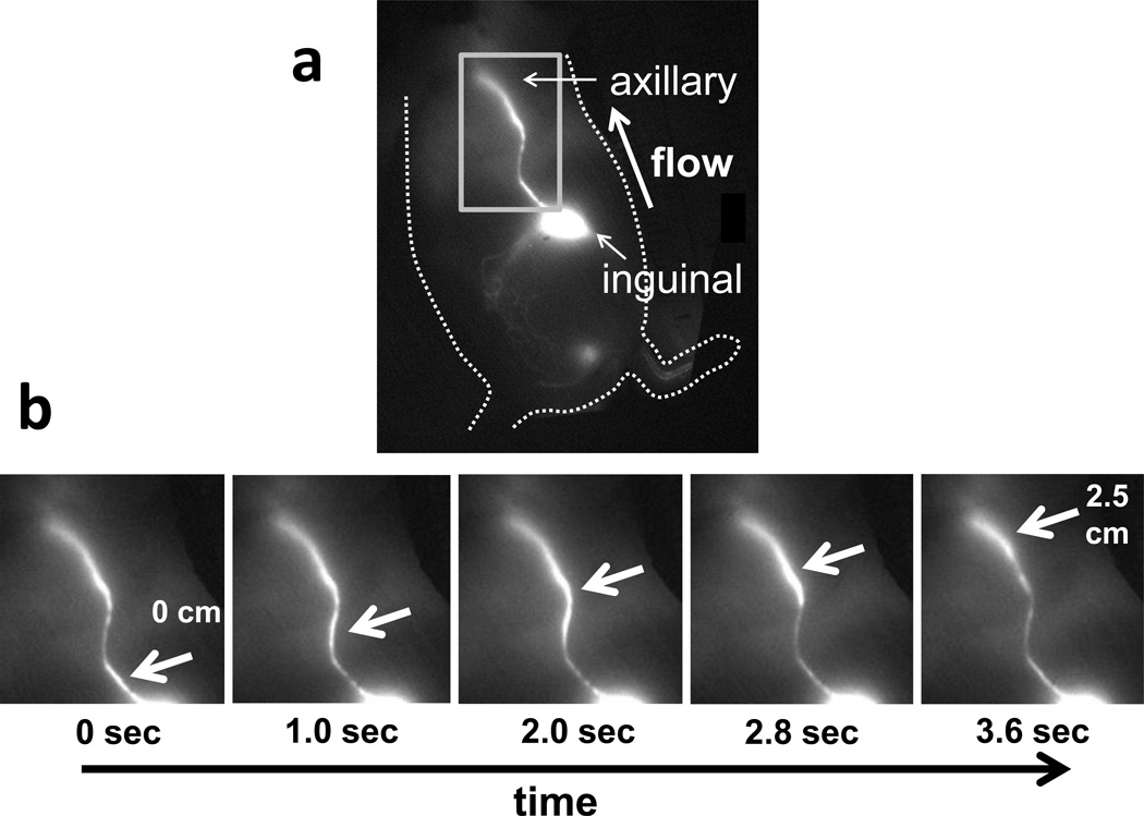
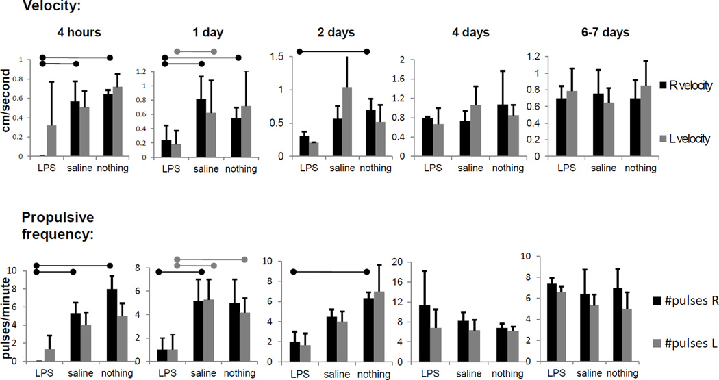
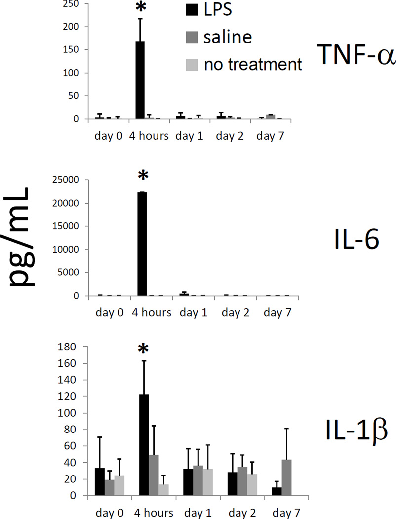
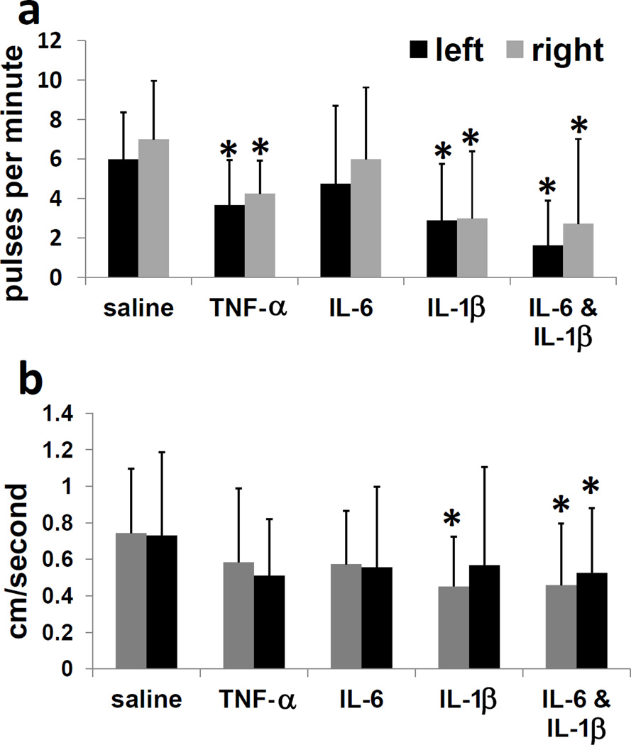
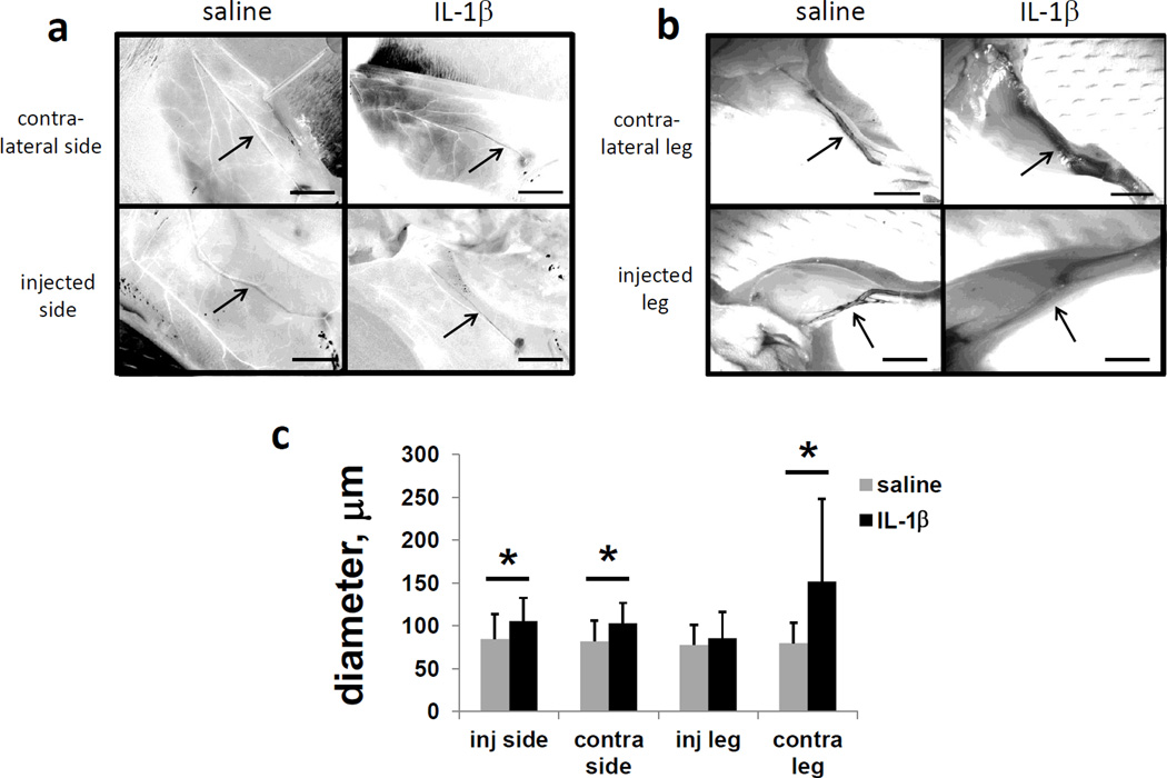
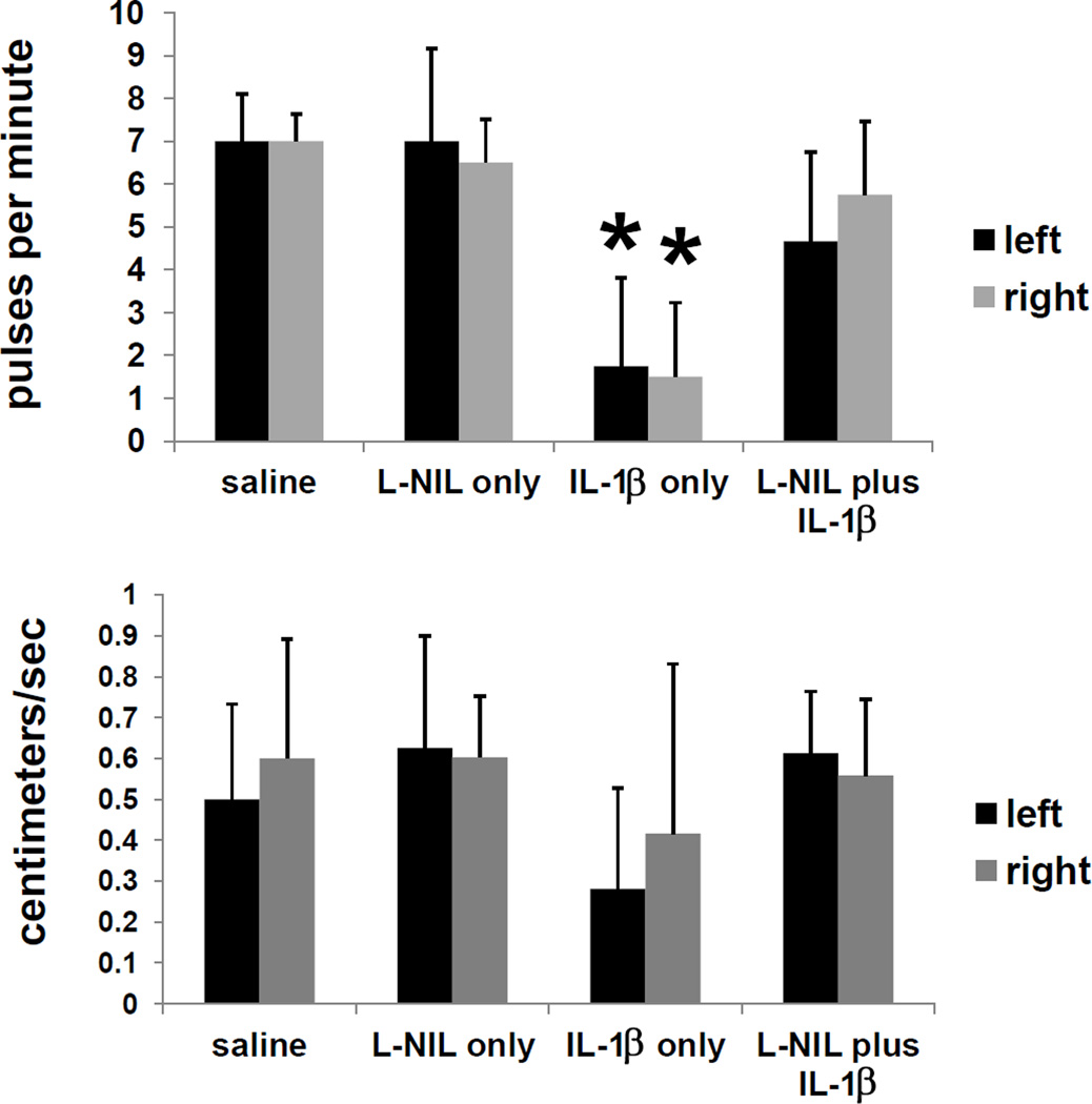
The response of the lymphatic system to inflammatory insult and infection is not completely understood. Using a near-infrared fluorescence (NIRF) imaging system to noninvasively document propulsive function, we noted the short-term cessation of murine lymphatic propulsion as early as 4h following LPS injection. Notably, the effects were systemic, displaying bilateral lymphatic pumping cessation after a unilateral insult. Furthermore, IL-1β, TNF-α, and IL-6, cytokines that were found to be elevated in serum during lymphatic pumping cessation, were shown separately to acutely and systemically decrease lymphatic pulsing frequency and velocity following intradermal administration. Surprisingly, marked lymphatic vessel dilation and leakiness were noted in limbs contralateral to IL-1β intradermal administration, but not in ipsilateral limbs. The effects of IL-1β on lymphatic pumping were abated by pre-treatment with an inhibitor of inducible nitric oxide synthase, L-NIL (N-iminoethyl-L-lysine). The results suggest that lymphatic propulsion is systemically impaired within 4h of acute inflammatory insult, and that some cytokines are major effectors of lymphatic pumping cessation through nitric oxide-mediated mechanisms. These findings may help in understanding the actions of cytokines as mediators of lymphatic function in inflammatory and infectious states.

Keywords: CD14/TLR4/MD2; CD14/Toll-like receptor 4/MD2; ICG; IFN-γ; IL; Inflammation; Interleukin-1 beta; Interleukin-6; L-NIL; LECs; LPS; Lymphatic; MCP-1/CCL2; N-iminoethyl-L-lysine; NIRF; T helper 1-type; Th1-type; Tumor necrosis factor-alpha; iNOS; indocyanine green; inducible nitric oxide; interferon-gamma; interleukin; lipopolysaccharide; lymphatic endothelial cells; monocyte chemoattractant protein-1/chemokine (C–C motif) ligand 2; near-infrared fluorescence imaging.


Figures

Figure 1
Figure 1
(a) Fluorescent image of side of mouse, displaying inguinal-to-axillary collecting lymphatic vessel and direction of normal flow, and (b) typical images of lymphatic movement in a side view of a mouse. Panels show sequential images of rectangular area, with arrows pointing to lymph packet/bolus moving from the inguinal lymph node through a collecting lymphatic vessel up to the axillary region.
Figure 2
Figure 2
Lymphatic function decreased systemically and acutely in response to LPS administration. Pulsatile velocity, or centimeters per second, and propulsive frequency, or pulses per minute, in right-side and left-side inguinal-to-axillary lymphatic vessels at 4 hours, 1 day, 2 days, 4 days, and 7 days after intradermal LPS injection (100 µg/mouse), saline injection (20 µl) or no injection in dorsal aspect of right hind paw. n=4–7 mice per group. Bars over column graphs indicate p<0.05.
Figure 3
Figure 3
TNF-α, IL-6, and IL-1β levels increase within 4 hours of LPS administration. TNF-α, IL-6, and IL-1β levels in mouse serum, as measured by Milliplex kit, at 0 hours, 4 hours, 1 day, 2 days, and 7 days after intradermal LPS (100 µg/mouse), saline, or no injection in dorsal aspect of right hind paw. n=5 mice per group. *=p<0.05 compared to saline or no injection.
Figure 4
Figure 4
Lymphatic function decreases systemically and acutely in response to administration of TNF-α, IL-6, and IL-1β. (a) Propulsive frequency, or pulses per minute, and (b) pulsatile velocity, or centimeters per second, in right-side and left-side inguinal-to-axillary lymphatic vessels at 4 hours after intradermal TNF-α (2 ug/mouse), IL-6 (50 ng/mouse), IL-1β (1 ug/mouse), or IL-6 plus IL-1β injection in dorsal aspect of right hind paw. n=10–16 mice per group. *=p<0.05 compared to saline.
Figure 5
Figure 5
Hindlimb lymphatic vessels contralateral to IL-1β injection are dilated/leaky. Intravital images of inguinal-to-axillary and hindlimb lymphatic vessels stained with 1% Evan’s blue dye injected i.d. in paws and at base of tail 4 hours after injection of saline or IL-β(1 ug/mouse) in the dorsal aspect of the right or left hind paw. (a) Inguinal-to-axillary lymphatic vessels, (b) hindlimb lymphatic vessels between paw and popliteal lymph nodes, and (c) average vessel diameters. After image conversion to HSB stack, lymphatic vessels appear black, and blood vessels appear white. n=10 mice per group. Scale bars=5 mm. *=p<0.05. Images are representative of 10 mice per group.
Figure 6
Figure 6
Attenuation of the effects of IL-1β on lymphatic function by the iNOS inhibitor L-NIL. Pulsatile frequency and velocity in mice after iNOS inhibitor (L-NIL) treatment. Mice were injected with 200 µg (i.p.) L-NIL or saline, then IL-1β (1 µg/mouse, i.d. in dorsal aspect of right hind paw) or saline one hour later, and imaged with ICG four more hours later. n=6 mice per group. *=p<0.05.

Similar articles

Cited by

References

    1. Stedman TL. Stedman’s Medical Dictionary. 28 th. Baltimore, Maryland: Lippincott Williams and Wilkins; 2006.
    1. Alitalo K. The lymphatic vasculature in disease. Nat Med. 2011;17:1371–1380. - PubMed
    1. Wang Y, Oliver G. Current views on the function of the lymphatic vasculature in health and disease. Gene Dev. 2010;24:2115–2126. - PMC - PubMed
    1. Kwon S, Sevick-Muraca EM. Noninvasive quantitative imaging of lymph function in mice. Lymphat Res Biol. 2007;5:219–231. - PubMed
    1. Rasmussen JC, Tan I-C, Marshall MV, Adams KE, Kwon S, Fife CE, Maus EA, Smith LA, Covington KR, Sevick-Muraca EM. Human lymphatic architecture and dynamic transport imaged using near-infrared fluorescence. Transl Oncol. 2010;3:362–372. - PMC - PubMed

Publication types

MeSH terms