. 2013 Jul-Aug;5(4):523-32.
doi: 10.4161/mabs.24979. Epub 2013 May 29.

Deep mutational scanning of an antibody against epidermal growth factor receptor using mammalian cell display and massively parallel pyrosequencing

Affiliations

Deep mutational scanning of an antibody against epidermal growth factor receptor using mammalian cell display and massively parallel pyrosequencing

Charles M Forsyth et al. MAbs. 2013 Jul-Aug.

Abstract

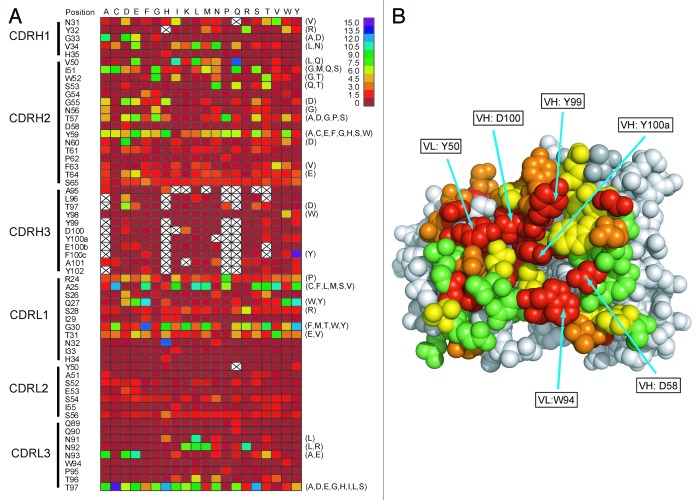
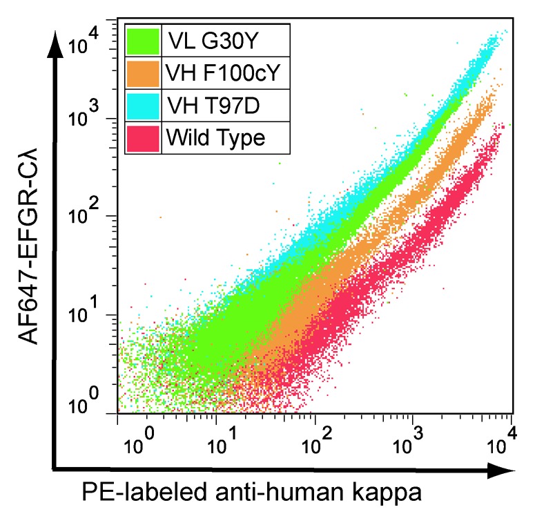
We developed a method for deep mutational scanning of antibody complementarity-determining regions (CDRs) that can determine in parallel the effect of every possible single amino acid CDR substitution on antigen binding. The method uses libraries of full length IgGs containing more than 1000 CDR point mutations displayed on mammalian cells, sorted by flow cytometry into subpopulations based on antigen affinity and analyzed by massively parallel pyrosequencing. Higher, lower and neutral affinity mutations are identified by their enrichment or depletion in the FACS subpopulations. We applied this method to a humanized version of the anti-epidermal growth factor receptor antibody cetuximab, generated a near comprehensive data set for 1060 point mutations that recapitulates previously determined structural and mutational data for these CDRs and identified 67 point mutations that increase affinity. The large-scale, comprehensive sequence-function data sets generated by this method should have broad utility for engineering properties such as antibody affinity and specificity and may advance theoretical understanding of antibody-antigen recognition.

Keywords: EGFR; antibody engineering; cetuximab; mammalian display; next generation sequencing.


Figures

None
Figure 1. Humanization of murine antibody 225 to create hu225. heavy (A) and light (B) variable domains of 225, hu225 and human acceptor frameworks 60P2 and NOV. CDRs (Kabat definition) are underlined in 225. Framework positions in hu225 where murine residues were retained are shown in bold and underlined. At VH position 12 double underlined and bold indicates where the infrequent Ile found in 60P2 was changed to Val as found in the human VH3 family consensus.
None
Figure 2. FACS sorting of 293c18 cells expressing surface-displayed hu225 and libraries. (A) wild type hu225 and (B-C) hu225 VH and VL libraries, respectively, were stained with AF647-EGFR-Cλ fusion protein and anti-human kappa PE (to normalize for IgG surface expression). Approximately 108 cells from each library were sorted on a MoFlo MLS instrument using high affinity binding (Bind) and expression (Exp) gates are indicated.
None
Figure 3. ER correlation for synonymous codon pairs. Scatter plot showing enrichment ratios for 398 synonymous NNT/NNG codon pairs encoding Ala, Gly, Leu, Pro, Arg, Ser, Thr and Val substitutions,. ER for the NNG codon is shown on the X axis, ER for the corresponding NNT codon is shown on the Y axis.
None
Figure 4. Structural analysis and positional tolerance to mutation. (A) Heatmap showing color coded ERs for each hu225 point mutation (where applicable data are average of multiple codons). To right of the heatmap in parentheses are indicated 67 higher affinity point mutations confirmed by FACS-based assay. Blank spaces indicate variants for which fewer than 10 total occurrences were found in the expression gate and which were eliminated from the analysis. (B) Average ER for all codon variants at each position color-coded and plotted onto the cetuximab Fab / EGFR crystal structure. Color code: average ER 0–0.125 (red), 0.125–0.75 (orange), 0.75–1.5 (yellow) and greater than 1.5 (green).
None
Figure 5. FACS analysis confirming higher affinity for selected hu225 variants. X axis = staining with anti-human kappa PE and Y axis = staining with AF647-EGFR-Cλ. A variant with vertical shift in relation to wild type indicates increased binding affinity.

References

    1. Carter PJ. Potent antibody therapeutics by design. Nat Rev Immunol. 2006;6:343–57. doi: 10.1038/nri1837. - DOI - PubMed
    1. Presta LG. Molecular engineering and design of therapeutic antibodies. Curr Opin Immunol. 2008;20:460–70. doi: 10.1016/j.coi.2008.06.012. - DOI - PubMed
    1. Metzker ML. Sequencing technologies - the next generation. Nat Rev Genet. 2010;11:31–46. doi: 10.1038/nrg2626. - DOI - PubMed
    1. Araya CL, Fowler DM. Deep mutational scanning: assessing protein function on a massive scale. Trends Biotechnol. 2011;29:435–42. doi: 10.1016/j.tibtech.2011.04.003. - DOI - PMC - PubMed
    1. Sato JD, Kawamoto T, Le AD, Mendelsohn J, Polikoff J, Sato GH. Biological effects in vitro of monoclonal antibodies to human epidermal growth factor receptors. Mol Biol Med. 1983;1:511–29. - PubMed

Publication types

MeSH terms