. 2013 Sep;27(9):1832-40.
doi: 10.1038/leu.2013.180. Epub 2013 Jun 14.

Toll-like receptor alterations in myelodysplastic syndrome

Affiliations

Toll-like receptor alterations in myelodysplastic syndrome

Y Wei et al. Leukemia. 2013 Sep.

Abstract

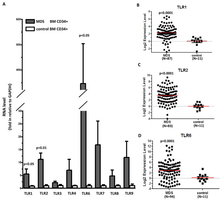
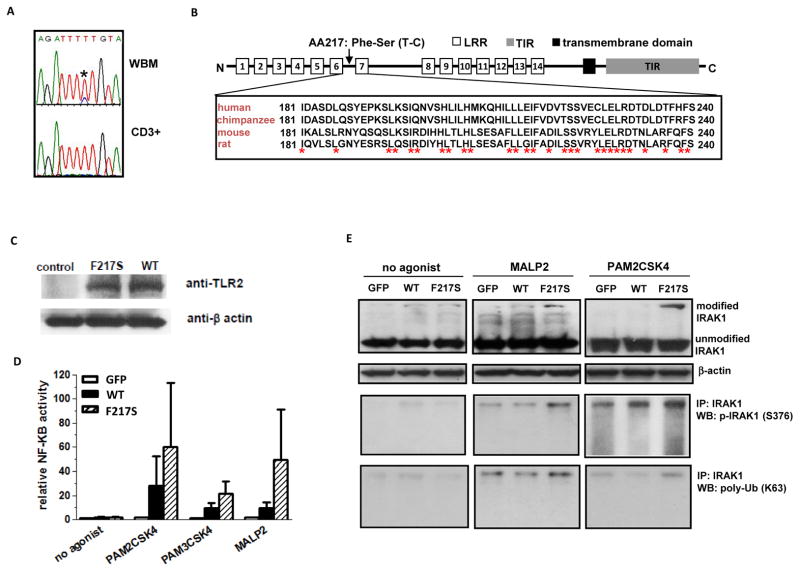
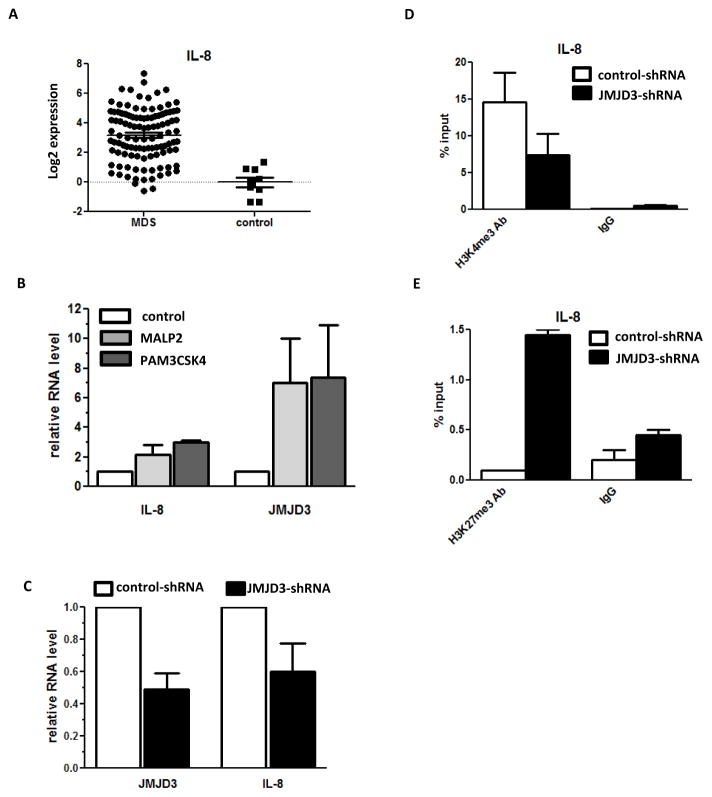
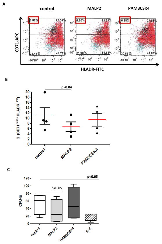
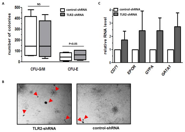
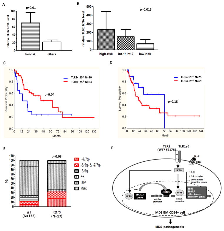
Recent studies have implicated the innate immunity system in the pathogenesis of myelodysplastic syndromes (MDS). Toll-like receptor (TLR) genes encode key innate immunity signal initiators. We recently identified multiple genes, known to be regulated by TLRs, to be overexpressed in MDS bone marrow (BM) CD34+ cells, and hypothesized that TLR signaling is abnormally activated in MDS. We analyzed a large cohort of MDS cases and identified TLR1, TLR2 and TLR6 to be significantly overexpressed in MDS BM CD34+ cells. Deep sequencing followed by Sanger resequencing of TLR1, TLR2, TLR4 and TLR6 genes uncovered a recurrent genetic variant, TLR2-F217S, in 11% of 149 patients. Functionally, TLR2-F217S results in enhanced activation of downstream signaling including NF-κB activity after TLR2 agonist treatment. In cultured primary BM CD34+ cells of normal donors, TLR2 agonists induced histone demethylase JMJD3 and interleukin-8 gene expression. Inhibition of TLR2 in BM CD34+ cells from patients with lower-risk MDS using short hairpin RNA resulted in increased erythroid colony formation. Finally, RNA expression levels of TLR2 and TLR6, as well as presence of TLR2-F217S, are associated with distinct prognosis and clinical characteristics. These findings indicate that TLR2-centered signaling is deregulated in MDS, and that its targeting may have potential therapeutic benefit in MDS.


Conflict of interest statement

CONFLICT OF INTEREST

None of the authors have any conflict of interest with the data presented here.

Figures

Figure 1
Figure 1. TLR RNA expression in primary MDS bone marrow CD34+ cells
(a) Q-RTPCR analysis of RNA expression of 8 TLR genes in MDS and control BM CD34+ cells. (b–d) Logarithmic representation of Q-RTPCR results of TLR1, 2, and 6 in MDS and control CD34+ cells. Numbers of samples with evaluable RNA expression are marked underneath of each graph.
Figure 2
Figure 2. Mutational analysis and identification of TLR2-F217S in MDS
(a) Representative Sanger sequencing traces showing nucleotides around coding region of TLR2-F217 in whole bone marrow (WBM) DNA (Top) and CD3+ genomic DNA (Bottom) of one MDS sample. (b) Schema of TLR2 gene and sequence alignments between human and other species. (c) Western blots of GFP fusion protein expression of TLR2 WT and F217S in 293T cells. (d) Luciferase analysis of activation of NF-kB by WT and F217S TLR2 in transfected 293T cells. Cells were exposed to either no agonist or to PAM2CSK4, PAM3CSK4 or MALP2 known TLR2 agonists. Pooled data from three separate experiments. (e) Western blot characterization of IRAK1 modification in 293T cells transfected with either WT or F217S TLR2 and treated with no agonist, MALP2 or PAM2CSK4. Top, Western blot analysis of IRAK1; Middle, IRAK1 immunoprecipitation followed by phospho-IRAK1 Western blot; Bottom, IRAK1 immunoprecipitation followed by poly-ubiquitin (K63).
Figure 3
Figure 3. Activation of IL-8 and JMJD3 gene expression by TLR2 signaling in primary BM CD34+ cells
(a) Logarithmic representation of Q-RTPCR result of IL-8 gene in MDS and control CD34+ cells. (b) Q-RTPCR analysis of IL-8 and JMJD3 RNA expression in cultured BM CD34+ cells after treatment with TLR2 agonist MALP2 and PAM3CSK4. (c) Q-RTPCR analysis of IL-8 RNA expression in OCI-AML3 cells after JMJD3 knock-down. (d) H3K4me3 CHIP-PCR analysis of IL-8 promoter in the OCI-AML3 cells after JMJD3 knock-down. (e) H3K27me3 CHIP-PCR analysis of IL-8 promoter in OCI- AML3 cells after JMJD3 knock-down.
Figure 4
Figure 4. Effect of TLR2 activation in in vitro cultured primary normal bone marrow CD34+ cells
(a) Flow cytometry analysis of normal CD34+ cells after being treated with MALP2 or PAM3CSK4 for 48 hours. Compared to control, treatment resulted in a decreased percentage of early erythroid progenitor cells marked with CD71+ and HLADR-. (b) Summary of flow cytometry analysis of CD71+ and HLADR- cells in primary BM CD34+ cells of four different donors. (c) Methocult medium supported colony formation assays revealed the decreased formation of erythroid colonies (CFU-E) from primary BM CD34+ cells treated by TLR2 agonist MALP2, PAM3CSK4 or IL-8.
Figure 5
Figure 5. Effect of TLR2 shRNA transduction in cultured MDS bone marrow CD34+ cells
(a) Numbers of myeloid colonies (CFU-G/M) and erythroid colonies (CFU-E) formed in methocult culture two weeks after transduction of TLR2-shRNA and control shRNA in BM CD34+ cells isolated from patients with lower-risk MDS (low-risk and intermediate-1). (b) Representative microphotographs of colonies formed in methocult plates after transduction of TLR2-shRNA (Left panel) and control shRNA (Right panel). Red arrows point to CFU-E. (c) Q-RTPCR analysis of the RNA levels of CD71, EPOR, GYPA and GATA1 in cells collected from total colonies after shRNA transduction and methocult assays.
Figure 6
Figure 6. Clinical analysis and proposed model of TLR2 mediated innate immunity signaling in MDS
(a) Higher TLR2 expression level in low-risk patients compared to others. (b) Distinct TLR6 expression levels in high-risk, intermediate-1 and 2, and low-risk patients. (c–d) Correlation of TLR2 and 6 with overall survival of patients. (e) Patients with TLR2-F217S are with higher frequency of 7-/7q-. (f) Proposed model of a potential TLR2-JMJD3-IL8 signaling axis is abnormally activated in BM CD34+ cells of MDS. This abnormal activation includes overexpression of TLR1, 2 and 6, as well as genetic alteration (F217S) of TLR2. This signal axis leads to consequent activation of JMJD3 and IL-8. JMJD3 can also positively regulate both genes of IL8 and its receptor in MDS BM CD34+ cells. Impact of this innate immunity signaling contributes to the pathogenesis of MDS.

References

    1. Tefferi A, Vardiman JW. Myelodysplastic syndromes. The New England journal of medicine. 2009;361(19):1872–85. - PubMed
    1. Wei Y, Chen R, Dimicoli S, Bueso-Ramos C, Neuberg D, Pierce S, et al. Global H3K4me3 genome mapping reveals alterations of innate immunity signaling and overexpression of JMJD3 in human myelodysplastic syndrome CD34+ cells. Leukemia: official journal of the Leukemia Society of America Leukemia Research Fund, U.K. 2013 - PMC - PubMed
    1. Akira S, Uematsu S, Takeuchi O. Pathogen recognition and innate immunity. Cell. 2006;124(4):783–801. - PubMed
    1. De Santa F, Totaro MG, Prosperini E, Notarbartolo S, Testa G, Natoli G. The histone H3 lysine-27 demethylase Jmjd3 links inflammation to inhibition of polycomb-mediated gene silencing. Cell. 2007;130(6):1083–94. - PubMed
    1. Li H, Durbin R. Fast and accurate short read alignment with Burrows-Wheeler transform. Bioinformatics. 2009;25(14):1754–60. - PMC - PubMed

Publication types

MeSH terms