. 2013 Dec;31(12):2833-41.
doi: 10.1002/stem.1445.

Brief report: VGLL4 is a novel regulator of survival in human embryonic stem cells

Affiliations

Brief report: VGLL4 is a novel regulator of survival in human embryonic stem cells

Adriana Tajonar et al. Stem Cells. 2013 Dec.

Abstract

Human embryonic stem cells (hESCs) are maintained in a self-renewing state by an interconnected network of mechanisms that sustain pluripotency, promote proliferation and survival, and prevent differentiation. We sought to find novel genes that could contribute to one or more of these processes using a gain-of-function screen of a large collection of human open reading frames. We identified Vestigial-like 4 (VGLL4), a cotranscriptional regulator with no previously described function in hESCs, as a positive regulator of survival in hESCs. Specifically, VGLL4 overexpression in hESCs significantly decreases cell death in response to dissociation stress. Additionally, VGLL4 overexpression enhances hESC colony formation from single cells. These effects may be attributable, in part, to a decreased activity of initiator and effector caspases observed in the context of VGLL4 overexpression. Additionally, we show an interaction between VGLL4 and the Rho/Rock pathway, previously implicated in hESC survival. This study introduces a novel gain-of-function approach for studying hESC maintenance and presents VGLL4 as a previously undescribed regulator of this process. Stem Cells 2013;31:2833-2841.

Keywords: Apoptosis; Pluripotent stem cells; Rho-associated kinases; VGLL4 protein.


Figures

Figure 1
Figure 1. Strategy for the identification of novel regulators of hESC maintenance
A) The hORFeome library is contained in a Gateway-adapted pHAGE lentiviral vector (top). The expression of the hemagglutinin (HA)-tagged ORF is driven by an EF1α (elongation factor 1 alpha) promoter, and a puromycin resistance gene driven by a PGK (phosphoglycerate kinase) promoter facilitates selection of transduced cells. Primer-binding sites are indicated by black arrows. The system was validated by transducing hESCs with a GFP ORF and selecting with 2 μg/ml of puromycin two days after transduction (bottom). Bar = 500 μm. B) Validation of screening conditions. Cells transduced with a Nanog ORF were used as a positive control, cells transduced with a GFP ORF and wildtype cells were used as negative controls. Negative controls lost the characteristic hESC morphology after three weeks or 3 passages of TGFβ inhibitor (TGFβi) treatment. Bars = 500 μm. C) Cells transduced with GFP and treated with TGFβi for three weeks downregulate Tra1–60 whereas NANOG-transduced cells do not. D) hESCs transduced with lentiviruses containing approximately 8000 genes were subjected to three weeks of treatment with 10μM TGFβ inhibitor. Colonies with characteristic ES cell morphology after treatment were isolated manually and their DNA was sequenced to identify 75 preliminary candidates (Supplementary Information Table S1). MOI=Multiplicity of infection.
Figure 2
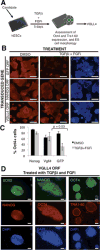
Figure 2. Verification of primary hits using a secondary verification assay
A) Preliminary candidates were verified by 5 days of treatment of transduced cells with with 20μM FGF inhibitor (FGFi) in addition to 10μM TGFβi. Following treatment, colony morphology and expression of hESC markers OCT4 and TRA 1–60 were analyzed. B) Cells transduced with VGLL4 or control genes were treated with vehicle control or with the inhibitor mix for 5 days and stained for OCT4 at the end of the treatment. Bars = 500 μm. C) Quantification of OCT4+ cells after treatment using Cellomics image acquisition and software. Error bars represent the standard deviation and p-values were obtained using a Student’s T-test. D) VGLL4-transduced cells treated with the inhibitor combination and stained for several hES cell-specific markers. Bars = 100 μm.
Figure 3
Figure 3. VGLL4 promotes survival and prevents apoptosis of hES cells through modulation of Caspase activation
A–D) AnnexinV and PI staining of VGLL4 or control hESCs maintained in self-renewal conditions (A) or treated with 500 μM EDTA (B) for 12–18 hours and analyzed by FACS. Live cells are APC, PI; apoptotic cells are APC+, PI, and dead cells are PI+. C, D) Flow cytometry data of cells processed as in (A, B) from 6 independent experiments. Error bars represent the standard deviation and the p-values were obtained with an unpaired Student’s T-test. E) Caspase 3/7 activity for VGLL4 and control hESCs after treatment with self-renewal or dissociation conditions (500 μM EDTA) using an activity-dependent caspase luminescence assay. Relative luminescence units (RLU) represent the signal after subtracting the background signal. Error bars represent the standard deviation of triplicates. P-values were obtained using an unpaired Student’s T-test. F) Increased caspase activity upon VGLL4 knockdown. Caspase 3/7 activity for scrambled control (mir neg) and VGLL4 knockdown (V78) in self-renewal and dissociation conditions measured as described in (E). Error bars represent the standard deviation from triplicate samples. P-values were calculated using an unpaired Student’s T-test. G) Colony formation efficiency for 100 cells/well treated with the indicated concentration of Rock inhibitor. H) Model for mode of action of VGLL4. Rho signaling is activated upon disruption of E-cadherin cell-to-cell contacts resulting in apoptosis. VGLL4 could inhibit cell death by modulating the expression of apoptosis and cytoskeleton genes. Alternatively, VGLL4 could have a role in the cytoplasm independent from its role as a co-transcriptional regulator in the nucleus.

References

    1. Watanabe K, Ueno M, Kamiya D, Nishiyama A, Matsumura M, Wataya T, Takahashi JB, Nishikawa S, Muguruma K, Sasai Y. A ROCK inhibitor permits survival of dissociated human embryonic stem cells. Nat Biotechnol. 2007 Jun;25:681–6. - PubMed
    1. Chen G, Hou Z, Gulbranson DR, Thomson JA. Actin-myosin contractility is responsible for the reduced viability of dissociated human embryonic stem cells. Cell Stem Cell. 2010 Aug 6;7:240–8. - PMC - PubMed
    1. Ohgushi M, Matsumura M, Eiraku M, Murakami K, Aramaki T, Nishiyama A, Muguruma K, Nakano T, Suga H, Ueno M, Ishizaki T, Suemori H, Narumiya S, Niwa H, Sasai Y. Molecular pathway and cell state responsible for dissociation-induced apoptosis in human pluripotent stem cells. Cell Stem Cell. 2010 Aug 6;7:225–39. - PubMed
    1. Ohgushi M, Sasai Y. Lonely death dance of human pluripotent stem cells: ROCKing between metastable cell states. Trends in Cell Biology. 2011;21:274–282. - PubMed
    1. Murphy GJ, Mostoslavsky G, Kotton DN, Mulligan RC. Exogenous control of mammalian gene expression via modulation of translational termination. Nat Med. 2006;12:1093–1099. - PubMed