

PKCβ promotes vascular inflammation and acceleration of atherosclerosis in diabetic ApoE null mice
- PMID: 23766264
- PMCID: PMC3865290
- DOI: 10.1161/ATVBAHA.112.301113
PKCβ promotes vascular inflammation and acceleration of atherosclerosis in diabetic ApoE null mice
Abstract
Objective: Subjects with diabetes mellitus are at high risk for developing atherosclerosis through a variety of mechanisms. Because the metabolism of glucose results in production of activators of protein kinase C (PKC)β, it was logical to investigate the role of PKCβ in modulation of atherosclerosis in diabetes mellitus.
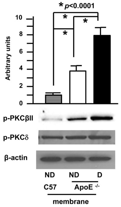
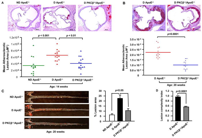
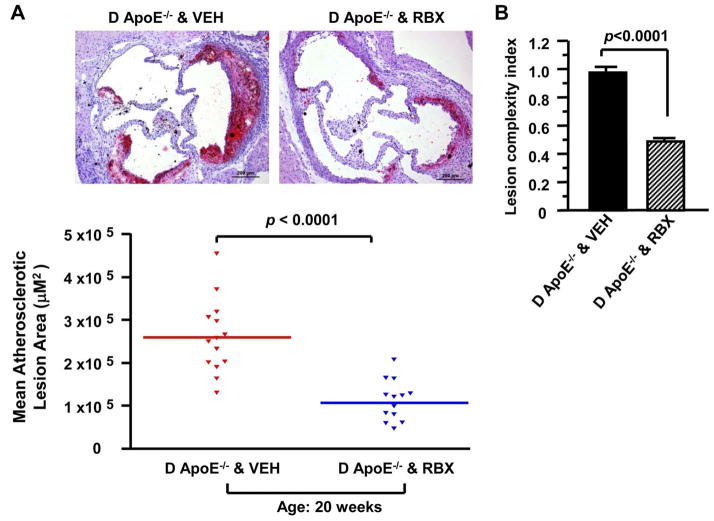
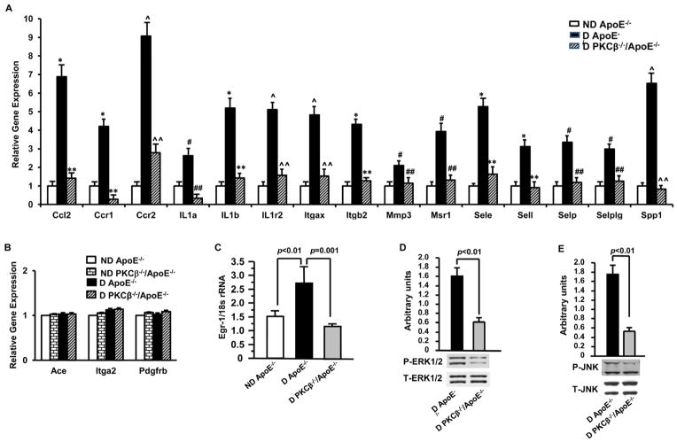
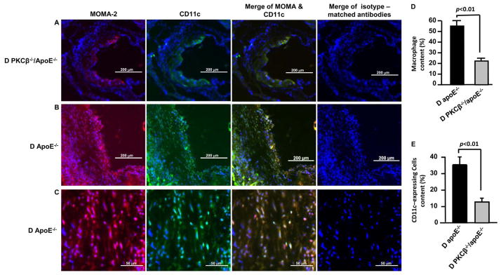
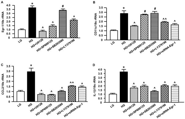
Approach and results: ApoE(-/-) and PKCβ(-/-)/ApoE(-/-) mice were rendered diabetic with streptozotocin. Quantification of atherosclerosis, gene expression profiling, or analysis of signaling molecules was performed on aortic sinus or aortas from diabetic mice. Diabetes mellitus-accelerated atherosclerosis increased the level of phosphorylated extracellular signal-regulated kinase 1/2 and Jun-N-terminus kinase mitogen-activated protein kinases and augmented vascular expression of inflammatory mediators, as well as increased monocyte/macrophage infiltration and CD11c(+) cells accumulation in diabetic ApoE(-/-) mice, processes that were diminished in diabetic PKCβ(-/-)/ApoE(-/-) mice. In addition, pharmacological inhibition of PKCβ reduced atherosclerotic lesion size in diabetic ApoE(-/-) mice. In vitro, the inhibitors of PKCβ and extracellular signal-regulated kinase 1/2, as well as small interfering RNA to Egr-1, significantly decreased high-glucose-induced expression of CD11c (integrin, alpha X 9 complement component 3 receptor 4 subunit]), chemokine (C-C motif) ligand 2, and interleukin-1β in U937 macrophages.
Conclusions: These data link enhanced activation of PKCβ to accelerated diabetic atherosclerosis via a mechanism that includes modulation of gene transcription and signal transduction in the vascular wall, processes that contribute to acceleration of vascular inflammation and atherosclerosis in diabetes mellitus. Our results uncover a novel role for PKCβ in modulating CD11c expression and inflammatory response of macrophages in the development of diabetic atherosclerosis. These findings support PKCβ activation as a potential therapeutic target for prevention and treatment of diabetic atherosclerosis.
Keywords: PKCβ; antigens, CD11c; atherosclerosis; diabetes mellitus; inflammation.
Figures
Comment in
-
Old suspect--new evidence: the role of PKCβ in diabetes mellitus-accelerated atherosclerosis.Arterioscler Thromb Vasc Biol. 2013 Aug;33(8):1737-8. doi: 10.1161/ATVBAHA.113.301922. Arterioscler Thromb Vasc Biol. 2013. PMID: 23864722 No abstract available.
References
-
- Ruderman NB, Gupta S, Sussman I. Hyperglycemia, diabetes and vascular disease: An overview. In: Ruderman N, Williamson J, Brownlee M, editors. Hyperglycemia, diabetes and vascular disease. New York, NY: Oxford University Press; 1992. pp. 3–20.
-
- Kannel WB, McGee DL. Diabetes and cardiovascular disease. The framingham study. JAMA. 1979;241:2035–2038. - PubMed
-
- Ferreiro JL, Angiolillo DJ. Diabetes and antiplatelet therapy in acute coronary syndrome. Circulation. 2011;123:798–813. - PubMed
-
- Luscher TF, Creager MA, Beckman JA, Cosentino F. Diabetes and vascular disease: Pathophysiology, clinical consequences, and medical therapy: Part ii. Circulation. 2003;108:1655–1661. - PubMed
-
- Haffner SM, Mykkanen L, Festa A, Burke JP, Stern MP. Insulin-resistant prediabetic subjects have more atherogenic risk factors than insulin-sensitive prediabetic subjects: Implications for preventing coronary heart disease during the prediabetic state. Circulation. 2000;101:975–980. - PubMed
Publication types
MeSH terms
Substances
Grants and funding
LinkOut - more resources
Full Text Sources
Other Literature Sources
Medical
Research Materials
Miscellaneous
