. 2013 Nov;94(5):963-70.
doi: 10.1189/jlb.0113051. Epub 2013 Jun 13.

Kupffer cells potentiate liver sinusoidal endothelial cell injury in sepsis by ligating programmed cell death ligand-1

Affiliations

Kupffer cells potentiate liver sinusoidal endothelial cell injury in sepsis by ligating programmed cell death ligand-1

Noelle A Hutchins et al. J Leukoc Biol. 2013 Nov.

Abstract

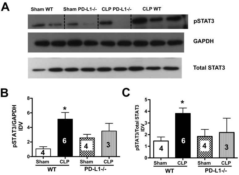
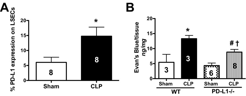
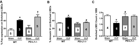
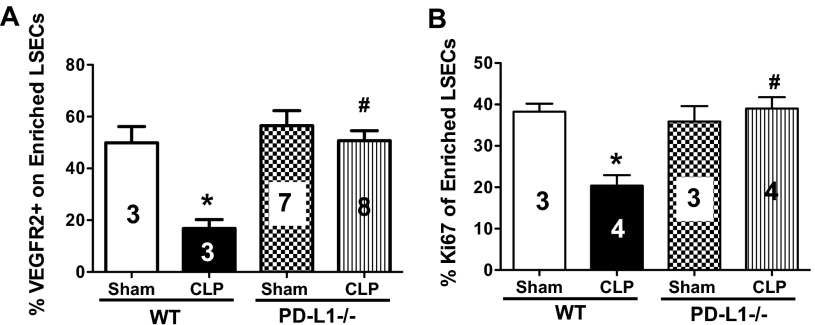
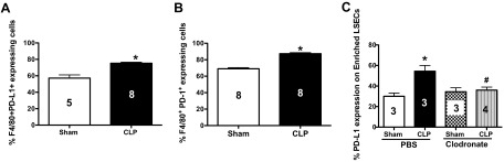
PD-1 and PD-L1 have been reported to provide peripheral tolerance by inhibiting TCR-mediated activation. We have reported that PD-L1-/- animals are protected from sepsis-induced mortality and immune suppression. Whereas studies indicate that LSECs normally express PD-L1, which is also thought to maintain local immune liver tolerance by ligating the receptor PD-1 on T lymphocytes, the role of PD-L1 in the septic liver remains unknown. Thus, we hypothesized initially that PD-L1 expression on LSECs protects them from sepsis-induced injury. We noted that the increased vascular permeability and pSTAT3 protein expression in whole liver from septic animals were attenuated in the absence of PD-L1. Isolated LSECs taken from septic animals, which exhibited increased cell death, declining cell numbers, reduced cellular proliferation, and VEGFR2 expression (an angiogenesis marker), also showed improved cell numbers, proliferation, and percent VEGFR2(+) levels in the absence of PD-L1. We also observed that sepsis induced an increase of liver F4/80(+)PD-1(+)-expressing KCs and increased PD-L1 expression on LSECs. Interestingly, PD-L1 expression levels on LSECs decreased when PD-1(+)-expressing KCs were depleted with clodronate liposomes. Contrary to our original hypothesis, we document here that increased interactions between PD-1(+) KCs and PD-L1(+) LSECs appear to lead to the decline of normal endothelial function-essential to sustain vascular integrity and prevent ALF. Importantly, we uncover an underappreciated pathological aspect of PD-1:PD-L1 ligation during inflammation that is independent of its normal, immune-suppressive activity.

Keywords: acute liver failure; coinhibitory molecules; hepatic endothelium; liver immune tolerance; septic inflammation.


Figures

Figure 1.
Figure 1.. PD-L1 modulates pSTAT3 expression in whole liver tissue in response to CLP.
(A) Representative Western blot, depicting cell lysates probed for pSTAT3, total STAT3, and GAPDH from Sham and CLP WT and PD-L1−/− animals. Dashed, black line identifies the sample. Bands were detected at 79, 86, and 37 kDa, respectively. (B and C) Densitometric analyses were performed on pooled Sham and CLP protein samples from WT and PD-L1−/− animals. Graphs indicate the densitometry quantification expressed as image density value (IDV) over total STAT3 and/or GAPDH. There is an increase of pSTAT3 expression (compared with GAPDH and total STAT3) in CLP WT livers versus Sham, n = 3–6, *P < 0.05. There is a declining trend of pSTAT3 expression in CLP PD-L1−/− livers: P > 0.05, CLP PD-L1−/− versus CLP WT; P > 0.05, CLP PD-L1−/− versus Sham PD-L1−/−. P values determined by a nonparametric one-way ANOVA. Data are expressed as mean ± sem. The numbers on the bars within this panel reflect the animals in each experimental group.
Figure 2.
Figure 2.. Increased PD-L1 expression on LSECs in response to CLP impacts liver tissue vascular permeability.
(A) LSECs up-regulate PD-L1 expression, 24 h post-CLP in WT animals, n = 8/group; *P < 0.05, CLP versus Sham WT, determined by a Mann-Whitney t-test. The numbers on the bar reflect the animals in each group. (B) There is more albumin protein leakage, as assessed by EBD, in liver tissue taken from CLP versus Sham WT animals, n = 3–8 group; *P < 0.05, CLP WT versus Sham WT. The increased EBD extravasation decreases in CLP PD-L1−/− animals, n = 3–8/group; #P < 0.05, CLP PD-L1−/− versus CLP WT; †P < 0.05, CLP PD-L1−/− versus Sham PD-L1−/−. P values were determined by a one-way ANOVA nonparametric test. All data in this panel are expressed as mean ± sem. The numbers on the bars within this panel reflect the animals in each experimental group.
Figure 3.
Figure 3.. Independent but synergetic effects of Fas and PD-L1 expression on CLP-induced mouse LSEC loss and apoptosis.
(A) Fas:FasL and PD-1:PD-L1 signaling events are independent of one another during sepsis. There is an increase of Fas expression on isolated LSECs from CLP WT and CLP PD-L1−/− animals versus their respective controls, n = 3–5; *P < 0.05, CLP versus Sham WT; †P < 0.05, CLP PD-L1−/− versus Sham PD-L1−/−. P values were determined by a nonparametric one-way ANOVA test. (B) There is an increase of Annexin V staining on isolated LSECs from CLP versus Sham WT animals, and this staining decreases in CLP PD-L1−/− LSECs, n = 3–6/group; *P < 0.05, CLP versus Sham WT; #P < 0.05, CLP PD-L1−/− versus CLP WT. P values determined by a nonparametric one-way ANOVA. (C) There is a decrease in LSEC number in CLP versus Sham WT animals, and these LSEC numbers are rescued in CLP PD-L1−/− animals, n = 4–8; *P < 0.05, CLP WT versus Sham WT; #P < 0.05, CLP PD-L1−/− versus CLP WT. P values determined by a nonparametric one-way ANOVA. All data in this panel are expressed as mean ± sem. The numbers on the bars within this panel reflect the animals in each experimental group.
Figure 4.
Figure 4.. PD-L1 expression on mouse LSECs modulates angiogenesis and cellular proliferation in response to CLP.
(A) There is a decrease of VEGFR2 expression on LSECs from CLP versus Sham WT animals, and this returns to Sham WT levels in LSECs from CLP PD-L1−/− animals, n = 3–8; *P < 0.05, CLP WT versus Sham WT; #P < 0.05, CLP PD-L1−/− versus CLP WT. P values were determined by a nonparametric one-way ANOVA test. (B) LSECs from CLP versus Sham WT animals proliferate less, n = 3–4; *P < 0.05, CLP WT versus Sham WT; there is greater proliferation in LSECs from CLP PD-L1−/− animals; #P < 0.05, CLP PD-L1−/− versus CLP WT (see Supplemental Fig. 1 for Ki67+ gating strategy). P values determined by a nonparametric one-way ANOVA. All data in this panel are expressed as mean ± sem. The numbers on the bars within this panel reflect the animals in each experimental group.
Figure 5.
Figure 5.. PD-1
+-expressing KCs ligate PD-L1 on LSECs during CLP. (A) There is an increase of liver F4/80+PD-L1+-expressing KCs following WT CLP at 24 h, CLP WT versus Sham WT, n = 5–8/group; *P < 0.05, determined by a nonparametric Mann-Whitney t-test. (B) There is an increase of liver F4/80+PD-1+-expressing KCs following WT CLP at 24 h versus Sham WT, n = 8/group; *P < 0.05, determined by a nonparametric Mann Whitney t-test. (C) There is an increase of PD-L1 expression on LSECs from CLP PBS control animals, n = 4–5/group; *P < 0.05, CLP PBS versus Sham PBS. PD-L1 expression levels on LSECs in CLP-KC-depleted animals (clodronate-treated) decreases; #P < 0.05, CLP-clodronate versus CLP PBS. P values were determined by a nonparametric one-way ANOVA test. All data in this panel are expressed as mean ± sem. The numbers on the bars within this panel reflect the animals in each experimental group.

References

    1. Angus D. C., Linde-Zwirble W. T., Lidicker J., Clermont G., Carcillo J., Pinsky M. R. (2001) Epidemiology of severe sepsis in the United States: analysis of incidence, outcome, and associated costs of care. Crit. Care Med. 29, 1303–1310 - PubMed
    1. Canabal J. M., Kramer D. J. (2008) Management of sepsis in patients with liver failure. Curr. Opin. Crit. Care 14, 189–197 - PubMed
    1. Aird W. C. (2003) The role of the endothelium in severe sepsis and multiple organ dysfunction syndrome. Blood 101, 3765–3777 - PubMed
    1. Hutchins N. A., Chung C. S., Borgerding J. N., Ayala C. A., Ayala A. (2013) Kupffer cells protect liver sinusoidal endothelial cells from Fas-dependent apoptosis in sepsis by down-regulating gp130. Am. J. Pathol. 182, 742–754 - PMC - PubMed
    1. Crispe I. N. (2009) The liver as a lymphoid organ. Annu. Rev. Immunol. 27, 147–163 - PubMed

Publication types