doi: 10.5489/cuaj.1219.
Genitourinary tuberculosis masquerading as a ureteral calculus
Affiliations
- PMID: 23766841
- PMCID: PMC3668412
- DOI: 10.5489/cuaj.1219
Item in Clipboard
Genitourinary tuberculosis masquerading as a ureteral calculus
Can Urol Assoc J.
2013 May-Jun.
Abstract
The genitourinary tract is a common extrapulmonary site of tuberculosis infection, yet remains a rare clinical entity in North America. We report the case of a 37-year-old man who presented for extracorporeal shock wave lithotripsy for a suspected ureteral stone on imaging. Further workup confirmed a diagnosis of genitourinary tuberculosis. Medical management was undertaken and, ultimately, nephrectomy performed. This case highlights the importance of maintaining a high index of clinical suspicion for genitourinary tuberculosis.
Figures
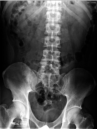
A kidney, ureter, bladder (KUB) radiograph demonstrating an oval density over the left hemipelvis.
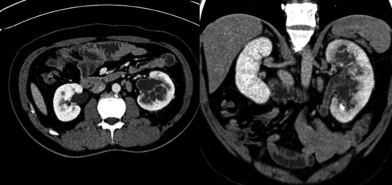
Axial (a) and coronal (b) abdominal computed tomography images showing small septated-appearing cystic spaces throughout the left renal parenchyma, left hydronephrosis, and calcifications.
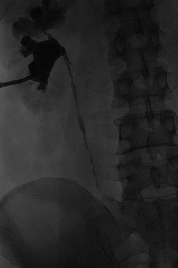
Left antegrade nephrostogram demonstrating scarring and dilatation of the upper pole with an abnormal infundibulum. The ureter demonstrates irregularity with a “beaded corkscrew” appearance.
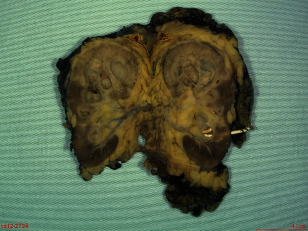
Gross pathology of the left nephrectomy specimen showing a 3.5-cm in diameter cavitary lesion filled with inflammatory exudate in the upper pole. The lesion involves the renal pelvis, which extended into the ureter distally occluding of the lumen.
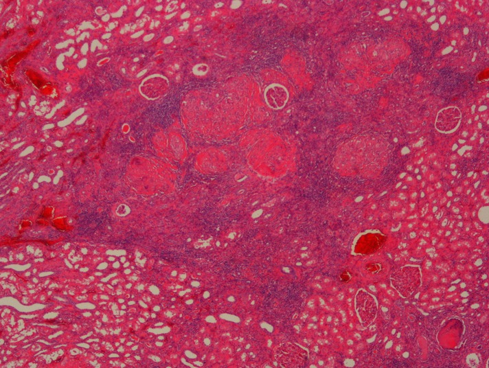
Left nephrectomy specimen micrograph showed numerous foci of necrotizing granulomatous inflammation in the renal cortex.
References
LinkOut - more resources
Full Text Sources
Other Literature Sources