Case Reports
. 2012 Jul;2(2):138-41.
doi: 10.4103/2229-5070.105183.

A rare case of disseminated cysticercosis

Affiliations
Case Reports

A rare case of disseminated cysticercosis

Kalpana A Bothale et al. Trop Parasitol. 2012 Jul.

Abstract

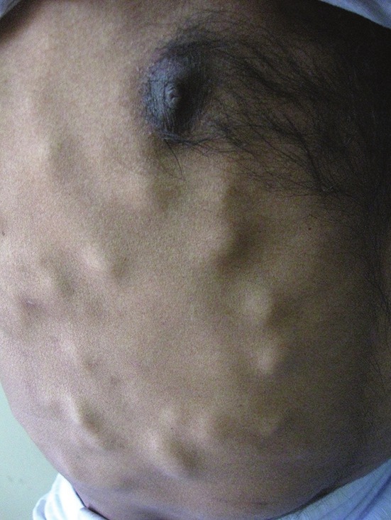
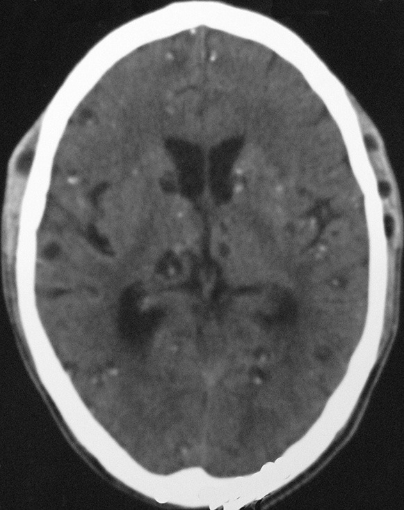
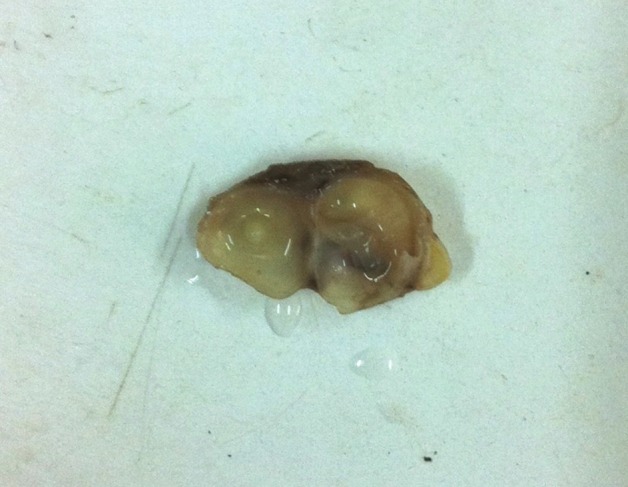
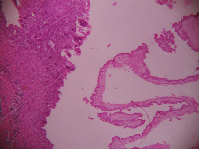
Cysticercosis is a common tropical disease. One of the uncommon manifestations and a rare complication is its disseminated form (DCC). Neurocysticercosis (NCC) is the common parasitic disease of the central nervous system. Human cysticercosis is caused by the dissemination of the embryo of Taenia solium in the intestine via the hepatoportal system to the tissues and organs of the body. The organs most commonly affected are the subcutaneous tissues, skeletal muscles, lungs, brain, eyes, liver, and occasionally the heart, thyroid, and pancreas. Widespread dissemination of the cysticerci can result in the involvement of almost any organ in the body. We report here a case of a 36-year-old-male with disseminated cysticercosis. He visited our hospital with symptoms of multiple palpable nodules, dementia, and confusion. After the investigations he was diagnosed with disseminated cysticercosis involving the brain, subcutaneous tissues all over the body, and the skeletal muscles. The patient was initially treated with Albendazole in a private hospital, but there was no response. Then he was treated with Praziquantel and steroids.

Keywords: Disseminated cysticercosis; Neurocysticercosis; cysticercosis.


Conflict of interest statement

Conflict of Interest: None declared

Figures

Figure 1
Figure 1
Clinical photograph showing multiple, subcutaneous, nodular lesions over the forearm
Figure 2
Figure 2
Clinical photograph showing multiple nodular lesions over chest wall and abdomen
Figure 3
Figure 3
CT scan of the brain showing multiple scattered lesions of cysticercosis throughout the brain parenchyma, with some showing eccentric calcification
Figure 4
Figure 4
A biloculated, thin-walled cyst containing watery fluid and a tiny white area in the center
Figure 5
Figure 5
Histopathology of a subcutaneous forearm nodule, showing encysted larva of the cysticercus surrounded by a chronic inflammatory infiltrate and giant-cell reaction (H and E, × 100)

References

    1. Kumar A, Bhagwani DK, Sharma RK, Kavita, Sharma S, Datar S, et al. Disseminated cysticercosis. Indian Pediatr. 1996;33:337–9. - PubMed
    1. Bhalla A, Sood A, Sachdev A, Varma V. Disseminated cysticercosis: A case report and review of literature. J Med Case Rep. 2008;2:137. - PMC - PubMed
    1. Jain BK, Sankhe SS, Agrawal MD, Naphade PS. Disseminated cysticercosis with pulmonary and cardiac involvement. Indian J Radiol Imaging. 2010;20:310–3. - PMC - PubMed
    1. Krishnaswami CS. Case of cysticercuscellulose. Ind Med Gaz. 1912;27:43–4. - PubMed
    1. Bandyopadhyay D, Sen S. Disseminated cysticercosis with muscle hypertrophy. Indian J Dermatol. 2009;54:49–51. - PMC - PubMed

Publication types