

Regulation of muscle protein synthesis and the effects of catabolic states
- PMID: 23769967
- PMCID: PMC3759561
- DOI: 10.1016/j.biocel.2013.05.039
Regulation of muscle protein synthesis and the effects of catabolic states
Abstract
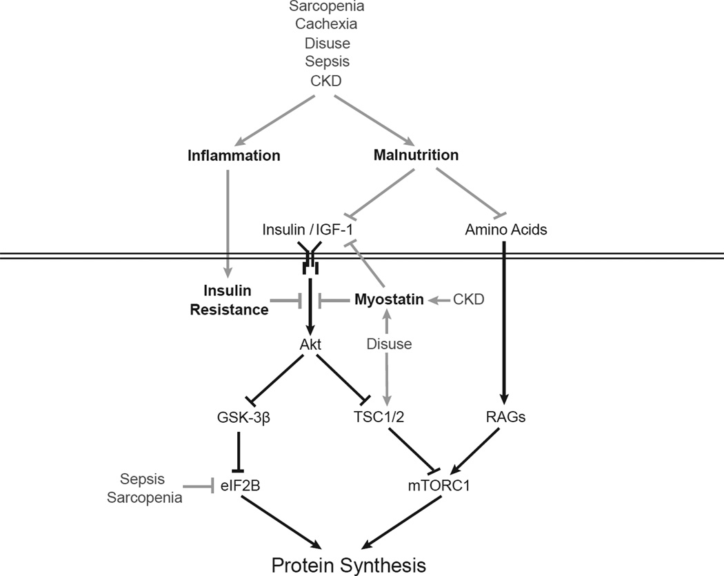
Protein synthesis and degradation are dynamically regulated processes that act in concert to control the accretion or loss of muscle mass. The present article focuses on the mechanisms involved in the impairment of protein synthesis that are associated with skeletal muscle atrophy. The vast majority of mechanisms known to regulate protein synthesis involve modulation of the initiation phase of mRNA translation, which comprises a series of reactions that result in the binding of initiator methionyl-tRNAi and mRNA to the 40S ribosomal subunit. The function of the proteins involved in both events has been shown to be repressed under atrophic conditions such as sepsis, cachexia, chronic kidney disease, sarcopenia, and disuse atrophy. The basis for the inhibition of protein synthesis under such conditions is likely to be multifactorial and includes insulin/insulin-like growth factor 1 resistance, pro-inflammatory cytokine expression, malnutrition, corticosteroids, and/or physical inactivity. The present article provides an overview of the existing literature regarding mechanisms and signaling pathways involved in the regulation of mRNA translation as they apply to skeletal muscle wasting, as well as the efficacy of potential clinical interventions such as nutrition and exercise in the maintenance of skeletal muscle protein synthesis under atrophic conditions. This article is part of a Directed Issue entitled: Molecular basis of muscle wasting.
Keywords: 3-phosphoinositol-dependent kinase 1; 4E-BP1; 5′-UTR; 5′-untranslated region; 70kDa ribosomal protein S6 kinase 1; AMP-activated protein kinase; AMPK; GCN2; GEF; GSK-3; HRI; ICU; IGF-1; IRES; Inflammation; Insulin resistance; LPS; Met-tRNA(i); Muscle atrophy; NF-κB; PDCD4; PDK1; PERK; PI3K; PKR; PKR-like endoplasmic reticulum-associated protein kinase; REDD; Rheb; TNF; TSC; double-stranded RNA-dependent protein kinase; eIF; eIF4E binding protein 1; eukaryotic initiation factor; general-control nonderepressible; glycogen synthase kinase-3; guanine nucleotide exchange factor; heme-regulated inhibitor; initiator methionyl-tRNA; insulin-like growth factor-1; intensive care unit; internal ribosome entry site; lipopolysaccharide; mRNA translation; mTOR; mTORC1; mechanistic target of rapamycin in complex 1; nuclear factor kappa-B; p70S6K1; phosphatidylinositol-4,5-bisphosphate 3-kinase; programmed cell death 4; ras homolog enriched in brain; regulated in DNA damage and development; tuberous sclerosis complex; tumor necrosis factor; uORF; upstream open reading frame.
Copyright © 2013 Elsevier Ltd. All rights reserved.
Figures
References
-
- Adey D, Kumar R, McCarthy JT, Nair KS. Reduced synthesis of muscle proteins in chronic renal failure. Am J Physiol Endocrinol Metab. 2000;278:E219–E225. - PubMed
-
- Allen DL, Linderman JK, Roy RR, Grindeland RE, Mukku V, Edgerton VR. Growth hormone/IGF-I and/or resistive exercise maintains myonuclear number in hindlimb unweighted muscles. J Appl Physiol. 1997;83:1857–1861. - PubMed
-
- Bailey JL, Zheng B, Hu Z, Price SR, Mitch WE. Chronic kidney disease causes defects in signaling through the insulin receptor substrate/phosphatidylinositol 3-kinase/Akt pathway: implications for muscle atrophy. J Am Soc Nephrol. 2006;17:1388–1394. - PubMed
-
- Bajotto G, Sato Y, Kitaura Y, Shimomura Y. Effect of branched-chain amino acid supplementation during unloading on regulatory components of protein synthesis in atrophied soleus muscles. Eur J Appl Physiol. 2011;111:1815–1828. - PubMed
Publication types
MeSH terms
Substances
Grants and funding
LinkOut - more resources
Full Text Sources
Other Literature Sources
Research Materials
Miscellaneous
