. 2013 Sep;176(1-2):292-7.
doi: 10.1016/j.virusres.2013.05.019. Epub 2013 Jun 13.

Porcine hemagglutinating encephalomyelitis virus induces apoptosis in a porcine kidney cell line via caspase-dependent pathways

Affiliations

Porcine hemagglutinating encephalomyelitis virus induces apoptosis in a porcine kidney cell line via caspase-dependent pathways

Yungang Lan et al. Virus Res. 2013 Sep.

Abstract

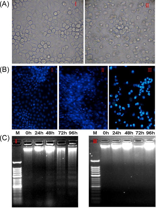
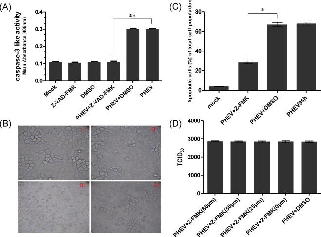
Porcine hemagglutinating encephalomyelitis is an acute, highly contagious disease in piglets that is caused by the porcine hemagglutinating encephalomyelitis virus (PHEV). However, the pathogenesis of PHEV and the relationship between PHEV and the host cells are not fully understood. In this study, we investigated whether the PHEV-induced cytopathic effect (CPE) was caused by apoptosis. Replication of PHEV in a porcine kidney-derived cell line (PK-15 cells) caused an extensive CPE, leading to the destruction of the entire monolayer and the death of the infected cells. Staining with Hoechst 33,342 revealed morphological changes in the nuclei and chromatin fragmentation. In addition, PHEV caused DNA fragmentation detectable by agarose gel electrophoresis 48h post-infection, increasing with the incubation time. The percentage of apoptotic cells increased with the incubation time and reached a maximum at 96h post-infection, as determined using flow cytometry and fluorescence microscopy of cells that were stained with annexin V-FITC and propidium iodide (PI). Moreover, as is commonly observed for coronavirus infections of other animals, the activities of the effecter caspase, caspase-3, and the initiator caspases, caspase-8 and caspase-9, which are representative factors in the death receptor-mediated apoptotic pathway and the mitochondrial apoptotic pathway, respectively, were increased in PHEV-infected PK-15 cells. Moreover, the tripeptide pan-ICE (caspase) inhibitor Z-VAD-FMK blocked PHEV-induced apoptosis but did not have an effect on virus production by 96h post-infection. These results suggested that PHEV induces apoptosis in PK-15 cells via a caspase-dependent pathway. Apoptotic death of infected cells is detrimental to animals because it causes cell and tissue destruction. Although the pathological characteristics of PHEV are largely unknown, apoptosis may be the pathological basis of the lesions resulting from PHEV infection.

Keywords: Apoptosis; Caspase; PK-15 cells; Procine haemagglutinating encephalomyelitis virus.


Figures

Fig. 1
Fig. 1
PHEV induces apoptosis in PK-15 cells. (A) Micrographs of mock-infected and PHEV infected PK-15 cells at 96 h post-infection. In contrast to mock-infected cells (I), PHEV-infected cells exhibit clear signs of shrinkage, rounding and detachment (II). (B) Cells were mock- or PHEV infected, and both adherent and non-adherent cells were isolated at 96 h post-infection, ethanol-fixed, stained with Hoechst 33,342, and viewed using a fluorescence microscope. Hoechst, nuclear staining. I: mock-infected cells, II: adherent infected cells, III: non-adherent infected cells. (C) Agarose gel electrophoresis of the DNA from PHEV-infected cells. DNA fragmentation was observed at and after 48 h post-infection, but was prominent at 72 and 96 h post-infection. I: infected cells M: 50-bp DNA marker, 0–96: 0, 24, 48, 72, and 96 h post-infection, II: mock-infected cells M: 50 bp DNA marker, 0–96: 0, 24, 48, 72, and 96 h post-infection. Cell images were captured at the conclusion of the study. The experiment was performed in triplicate and repeated three times.
Fig. 2
Fig. 2
Apoptotic PHEV-infected PK-15 cells stained with annexin V-FITC/PI were observed by fluorescence microscopy and analyzed by flow cytometry. (A) The samples were analyzed for green fluorescence (FITC) and red fluorescence (PI). Different labeling patterns of the PHEV-infected cells: early apoptotic cells, annexin V-FITC-positive and PI-negative cells (I); necrotic or late apoptotic cells, both annexin V-FITC- and PI-positive (II) cells; dead cells, Annexin V-FITC-negative and PI-positive cells (III). (B) The infected and mock-infected cells were harvested, stained with annexin V-FITC and PI, and analyzed by flow cytometry. Scatter plots of the annexin V-FITC/PI flow cytometry results of a representative experiment are presented below the graphs. The lower right quadrants represent cells in the early stage of apoptosis. The upper right quadrants represent cells in the late stage of apoptosis or necrotic cells. I-III: early and late apoptotic cells at 0, 72, and 96 h post-infection. (C) The data are expressed as the percentage of annexin V-FITC-positive cells (apoptotic cells) at different times. (two-way ANOVA with Bonferroni posttests *P < 0.05, **P < 0.01,***P < 0.001 compared with the data for mock-infected cells).
Fig. 3
Fig. 3
Kinetics of the activity of caspase-3 (A), caspase-8 (B) and caspase-9 (C) in PHEV-infected (&) and mock-infected (&) cells. Caspase 3, 8 and 9-like protease activity was expressed as the percentage of the activity of mock-infected cells 0 h (control), which was given a value of 100. The data represent the mean values ± SD. (two-way ANOVA with Bonferroni posttests *P < 0.05, **P < 0.01,***P < 0.001 compared with the values for mock-infected cells).
Fig. 4
Fig. 4
Effects of Z-VAD-FMK treatment on CPE, cell apoptosis and virus production in PHEV-infected PK-15 cells. (A) The data are expressed as the activity of caspase-3: mock-infected cells (Mock), Z-VAD-FMK treated cells (Z-VAD-FMK), DMSO treated cells (DMSO), cells infected with PHEV alone (PHEV), in the presence of DMSO (PHEV +DMSO) or Z-VAD-FMK (PHEV + Z-FMK-VAD). All determinations were done after 96 h of incubation. The data represent the mean values ± SD (unpaired-samples t-test **P < 0.01 compared with PHEV-infected cells). (B) Phase-contrast images of mock-infected (I), Z-VAD-FMK treated and infected (II), PHEV-infected (III), DMSO-treated and infected (IV), at an original magnification of 400. The experiment was performed in triplicate and repeated three times. (C) The data are expressed as the percentage of annexin V-FITC-positive (apoptotic cells) cells: mock-infected cells, cells treated with DMSO or Z-VAD-FMK, and cells infected with PHEV after 96 h of incubation (unpaired-samples t-test *P < 0.05 compared with the DMSO-treated and infected cells). (D) Virus titers at 96 h post-infection of PK-15 cells infected with PHEV alone (PHEV), in the presence of 0, 25, 50 and 80 μM Z-VAD-fmk (PHEV + Z-FMK), or DMSO (PHEV +DMSO).

References

    1. Andries K., Pensaert M. Propagation of hemagglutinating encephalomyelitis virus in porcine cell cultures. Zentralbl Veterinarmed B. 1980;27:280–290. - PMC - PubMed
    1. Arends M.J., Wyllie A.H. Apoptosis: mechanisms and roles in pathology. International Review of Experimental Pathology. 1991;32:223–254. - PubMed
    1. Cartwright S.F., Lucas M., Cavill J.P., Gush A.F., Blandford T.B. Vomiting and wasting disease of piglets. Veterinary Record. 1969;84:175–176. - PubMed
    1. Chen S., Cheng A.C., Wang M.S., Peng X. Detection of apoptosis induced by new type gosling viral enteritis virus in vitro through fluorescein annexin V-FITC/PI double labeling. World Journal of Gastroenterology. 2008;14:2174–2178. - PMC - PubMed
    1. Clarke P., Beckham J.D., Leser J.S., Hoyt C.C., Tyler K.L. Fas-mediated apoptotic signaling in the mouse brain following reovirus infection. Journal of Virology. 2009;83:6161–6170. - PMC - PubMed

Publication types