. 2013 Jun 27;38(6):1164-75.
doi: 10.1016/j.immuni.2013.02.023. Epub 2013 Jun 13.

Endocytosis and recycling of immune complexes by follicular dendritic cells enhances B cell antigen binding and activation

Affiliations

Endocytosis and recycling of immune complexes by follicular dendritic cells enhances B cell antigen binding and activation

Balthasar A Heesters et al. Immunity. .

Abstract

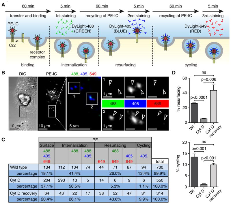
Stromal-derived follicular dendritic cells (FDCs) are a major reservoir for antigen that are essential for formation of germinal centers, the site where memory and effector B cells differentiate. A long-standing question is how FDCs retain antigen in its native form for extended periods and how they display it to specific B cells. Here we found that FDCs acquired complement-coated immune complexes (ICs) from noncognate B cells via complement receptors 1 and 2 (CD35 and CD21, respectively) and rapidly internalized them by an actin-dependent pathway. ICs were retained intact within a nondegradative cycling compartment and were displayed periodically on the cell surface where they were accessible to antigen-specific B cells. This would explain how antigens are protected from damage and retained over long periods of time, while remaining accessible for B cells.


Figures

Figure 1
Figure 1
(see also Figure S1, S2 and Movie S1, S2, S3). Acquisition of IC by FDC. (A) (left panel) Snap shot taken from multiphoton-intravital imaging of the popliteal LN in a mouse passively immunized with anti-PE antibody and adoptively transferred with WT fluorescently labeled B cells (green). LN was imaged from 60 min to 120 min following PE (red) injection into the footpad showing B cells transfer PE-IC onto FDC (blue). (right panel) Series of higher magnification images over 5 min showing transfer of PE-IC (red) bound to a B cell (green) to an FDC (blue) (arrowheads). (B) Live-cell imaging of cultured FDC labeled with lipophilic DiO dye (green) and incubated with PE-IC bound non-cognate B cells. Images were generated every 60 seconds. Images show transfer of C3-opsonized PE-IC clustered on the B cell to the FDC and subsequent fragmentation on the FDC surface. (right panel) Schematic representation of transfer of PE-IC clusters from B cells onto FDC and their fragmentation upon contact with FDC. (C) (Left panel) Spinning disk confocal image of PE-IC (red) within FDC (green). (right panel) Representative Z-stack series projection through 30 μm at approximate 0.4 μm intervals identifies PE-IC (grey and arrowheads) within FDC (outlined).
Figure 2
Figure 2
Antigen uptake by FDC is actin dependent. (A) FDC were treated with 5 μM Cytochalasin D (Cyt D) or media for 30 min, washed and then incubated with C3-opsonized PE-IC-loaded non-cognate B cells for another 30 min. (B) Representative confocal images of fixed cells show PE-IC (red) uptake by FDC from B cells is diminished in Cyt D treated FDC. FDC were identified by FDC-M1 (green) and Cr2 (blue) staining. (C) Quantification of confocal images by Volocity software showing PE mean fluorescent intensity per cell (PE intensity/number of voxels) for each group. (D) Cr2 MFI is not altered by Cyt D treatment. Cumulative data of FDC culture from 3 mice over 2 independent experiments (minimum of 15 cells per group). Each symbol represents a single cell; horizontal bar is mean value. Statistics: p-values calculated by Student's t test; ns = not significant.
Figure 3
Figure 3
Antigen acquired by FDC resurfaces. (A) Schematic diagram of FDC loading and acid wash procedure and recovery. PE-IC appearing on the surface of the FDC after acid wash must come from inside the cell. FDC were loaded with C3-opsonized PE-IC, then either fixed, acid washed and fixed or acid washed, recovered for 30 min and then fixed. Cells were stained with anti-PE and anti-C3d antibody. (B) Quantification of the results shows efficient stripping of C3-opsonized PE-IC by acid wash and recovery of PE-IC on the surface after 30 min. C3d remains fixed on PE-IC during resurfacing. (C) Schematic representation of the experiment. FDC were loaded with TEL (turkey egg lysozyme)-PE-IC or PE-IC by non-cognate B cells in culture. FDC were acid washed to remove cell surface antigen and fixed with 1 % PFA or left in media alone to recover. MD4 Ig transgenic B cells (specific for TEL) were mixed with FDC for 2 or 4 hr in culture, then harvested, stained with B220 and analyzed by flow cytometry. (D) Results indicate efficient uptake of TEL-PE-IC by cognate MD4 B cells when cultured with live FDC but negligible uptake of PE-IC (without TEL) or TEL-PE-IC from FDC after acid wash and fixation. (E) MD4 B cells were cultured for an additional 20 hours before staining with anti-CD86 to assay for B cell activation. Results indicate cognate B cells express CD86 when exposed to specific antigen on FDC but negligible expression when cultured with FDC loaded with non-specific PE-IC. Statistics: p-values calculated by Student's t test; ns = not significant.
Figure 4
Figure 4
FDC display of antigen for acquisition by cognate B cells is actin dependent. (A) Non-cognate B cells bearing clusters of NP haptenated PE-IC were incubated with FDC culture for 2 hr. Non-cognate B cells were removed by washing and pulsed FDC were treated with media alone, 1 or 5 μM Cyt D for 30 min. Cyt D was removed and the FDC were incubated with a mix of WT (IgMb) and NP-specific B1-8 (IgMa) B cells for 30 min before harvest and analysis by flow-cytometry. (B) Gating scheme for NP-PE-IC bound by B1-8 B cells (B220+, IgMa+). Histogram shows uptake is specific for B1-8 (IgMa) B cells but not WT (IgMb) as expected. (C) Percent PE positive B1-8 B cells (left panel) and the MFI of NP-PE-IC bound by B1-8 B cells (right panel). Results indicate that display of specific antigen on FDC is actin dependent. Results shown are from 3 separate experiments with 3 mice each. (D) FDC were loaded with NP-PE-IC as described above and cultured for 60 min before removal of the FDC. Subsequently, B1-8 B cells were mixed with FDC supernatant or directly with FDC in fresh media for 30 min. (E) B1-8 cells were harvested and analyzed by flow cytometry as in B and C. Results show efficient acquisition of antigen by B1-8 B cells requires intact FDC. Results represent 3 separate FDC cultures derived from 2 sets of mice. Statistics: p-values calculated by Student's t test; ns = not significant.
Figure 5
Figure 5
(see also Figure S3). Cycling of IC is actin dependent. (A) Schematic representation of the experimental set-up for antibody cell surface staining: C3-opsonized PE-IC is transferred to FDC for 60 min before removal of B cells. FDC are incubated with DyLight488 labeled donkey anti-rabbit IgG (green) for 5 minutes to detect surface PE-IC. FDC are washed, IC are allowed to cycle and the process is repeated with two additional labeled donkey anti-rabbit IgGs, i.e. DyLight405 (blue) and DyLight649 (red) respectively. This results in several color combinations that indicate cycling or resurfacing. (B) Representative confocal image of FDC with cycling PE-IC. PE channel (grey value) was used to identify objects and then objects were scored for green (DyLight488, first staining), blue (DyLight405, second staining) or red (DyLight649, third staining). If the PE-IC object was blue only it was scored as cycling (Inset 1). If the object was blue and green it was scored as internalization (Inset 2). (C) Table of all possible color combinations, their interpretation and the raw cumulative data. This setup will result in an underestimation of the actual number of cycling PE-IC. (D) Bar graph showing percent PE-IC resurfacing (top panel). Bar graph showing percent PE-IC cycling (bottom panel). Statistics: p-values calculated by Student's t test; ns = not significant; data are represented as mean +/- SEM.
Figure 6
Figure 6
Antigen acquired by FDC in vivo is periodically cycled to the cell surface. (A) Schematic representation, mice were passively immunized with rabbit anti-PE on day -1, injected with PE on day 0, then irradiated on day 4 or 9 and FDC isolated on day 5 or 10. Alternatively, draining popliteal LNs (pLN) were harvested and cryo-preserved on day 10. (B) Sections of pLN show PE (red) localizing with Cr2 (blue) that marks the FDC within the B cell (B220, green) follicle at day 10 post-immunization. (C) Representative confocal images of FDC harvested at day 5 post-immunization with PE (red) and cultured in vitro until day 11. Cells were stained with antibody to Cr2 (blue). Quantification of images shows mean fluorescent intensity of PE per cell for FDC isolated from immunized and PBS control mice. Each symbol represents a single cell. (D) Representative confocal images of FDC isolated from immune mice at day 10 and cultured until day 16. FDC were either fixed, acid washed and fixed or acid washed, recovered for 30 min and then fixed. Cells were stained with anti-PE and anti-C3d antibody. (E) Quantification shows efficient acid wash and recovery of PE-IC (upper panel) and C3 (lower panel) on the surface after 30 min. C3d remains fixed on PE-IC during resurfacing. Results indicate C3-PE-IC recycle at day 16 after loading in vivo. Statistics: p-values calculated by Student's t test; ns = not significant.

Similar articles

Cited by

References

    1. Alimzhanov MB, Kuprash DV, Kosco-Vilbois MH, Luz A, Turetskaya RL, Tarakhovsky A, Rajewsky K, Nedospasov SA, Pfeffer K. Abnormal development of secondary lymphoid tissues in lymphotoxin beta-deficient mice. Proc Natl Acad Sci USA. 1997;94:9302–9307. - PMC - PubMed
    1. Bajenoff M, Germain RN. B cell follicle development remodels the conduit system and allows soluble antigen delivery to follicular dendritic cells. Blood. 2009;114:4989–4997. - PMC - PubMed
    1. Bergtold A, Desai DD, Gavhane A, Clynes R. Cell surface recycling of internalized antigen permits dendritic cell priming of B cells. Immunity. 2005;23:503–514. - PubMed
    1. Boulant S, Kural C, Zeeh JC, Ubelmann F, Kirchhausen T. Actin dynamics counteract membrane tension during clathrin-mediated endocytosis. Nat Cell Biol. 2011;13:1124–1131. - PMC - PubMed
    1. Burton GF, Kosco MH, Szakal AK, Tew JG. Iccosomes and the secondary antibody response. Immunology. 1991;73:271–276. - PMC - PubMed

Publication types