. 2013 Jul 10;79(1):153-66.
doi: 10.1016/j.neuron.2013.04.039. Epub 2013 Jun 13.

The thalamostriatal pathway and cholinergic control of goal-directed action: interlacing new with existing learning in the striatum

Affiliations

The thalamostriatal pathway and cholinergic control of goal-directed action: interlacing new with existing learning in the striatum

Laura A Bradfield et al. Neuron. .

Abstract

The capacity for goal-directed action depends on encoding specific action-outcome associations, a learning process mediated by the posterior dorsomedial striatum (pDMS). In a changing environment, plasticity has to remain flexible, requiring interference between new and existing learning to be minimized, yet it is not known how new and existing learning are interlaced in this way. Here we investigated the role of the thalamostriatal pathway linking the parafascicular thalamus (Pf) with cholinergic interneurons (CINs) in the pDMS in this process. Removing the excitatory input from Pf to the CINs was found to reduce the firing rate and intrinsic activity of these neurons and produced an enduring deficit in goal-directed learning after changes in the action-outcome contingency. Disconnection of the Pf-pDMS pathway produced similar behavioral effects. These data suggest that CINs reduce interference between new and existing learning, consistent with claims that the thalamostriatal pathway exerts state control over learning-related plasticity.


Figures

Figure 1
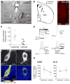
Figure 1. Parafascicular thalamic neurons directly and unilaterally project to dorsomedial striatal territories
(A) Fluorescent signal recorded in the parafscicular thalamic nucleus (Pf) 4 d after injection of retrograde tracer fluorogold in the DMS (inset). (B, C) Confocal higher-magnification images showing fluorogold fluorescence in Pf ipsilateral or contralateral to the DMS injection. D, dorsal; L, lateral.
Figure 2
Figure 2. Effect of parafascicular thalamic lesions on cholinergic interneurons of the dorsomedial striatum
(A) Rat Nissl-stained section showing unilateral NMDA excitotoxic lesions of the parafascicular nucleus (Pf). (B) Anatomical localization of randomly selected cholinergic interneurons from unlesioned and lesioned cerebral hemispheres. Each circle represents a single neuron sampled from electrophysiological recording. (C) Example of cholinergic interneuron labeled with biocytin in the DMS. (D) Cellular physiological characteristics of a recorded neuron under whole-cell patch-clamp. Current-voltage relationship recorded by stepping the cell to various hyperpolarizing membrane potentials (top-left panel). Under current-clamp configuration, whole-cell action potential (top-right panel) and depolarization-triggered action potential firing (bottom panels) were routinely sampled for comparisons with known CIN cellular characteristics. (E) Frequency distribution plot showing basal action potential firing in cholinergic interneurons of lesioned and unlesioned hemisections. (F) High-magnification confocal images showing p-Ser240–244-S6rp intensity in ChAT immunoreactive neurons of the DMS ipsilateral (lesioned) or contralateral (unlesioned) to the Pf lesion. A 16 pseudo-color palette (Lookup Table, LUT) highlights the intensity of p-S6rp fluorescence (display range: 0 – 4096). (G) Within-individuals quantification of p-Ser240–244-S6rp signal in ChAT immunoreactive neurons of dorsomedial (DMS) and dorsolateral (DLS) striatal territories ipsilateral or contralateral to the Pf lesion. In scatterplots each dot corresponds to one neuron; each color corresponds to a different animal.
Figure 3
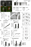
Figure 3. Parafascicular thalamus encodes changes in action-outcome contingencies
(A) Rats were trained on two lever press responses, R1 and R2, to earn pellets and sucrose outcomes (O1 and O2, counterbalanced). For test rats were sated for 1 hr on one outcome, i.e. O1 (1hr), prior to a choice test, R1 vs. R2. To induce new learning we used contingency degradation then reversal, which was tested using outcome devaluation and selective reinstatement tests. See supplemental methods for a full description of these procedures. (B) Rat Nissl-stained sections showing bilateral Sham or NMDA excitotoxic lesions of the parafascicular nucleus (Pf). (B–H) Mean or % baseline rate of lever pressing (± 1 SEM) during: (C) acquisition of initial action-outcome contingencies averaged over levers; (D) outcome devaluation testing; (E) acquisition of contingency degradation; (F) contingency degradation testing in extinction; (G) acquisition of the reversed contingencies; (H) outcome devaluation testing of the reversed contingencies; (I) reinstatement of reversed contingencies. (J) Minimal (black) and maximal (grey) extent of NMDA-induced excitotoxic lesions of the Pf.
Figure 4
Figure 4. Disconnection of the thalamo-striatal pathway confirms parafascicular involvement action-outcome learning in the posterior dorsomedial striatum
(A) Rat Nissl-stained sections showing anatomical disconnection through unilateral DMS and Pf NMDA excitotoxic lesions (sham and contralateral groups shown). (B–H) Mean or % baseline rate of lever pressing (±(1 SEM) during: (B) acquisition of initial action-outcome contingencies averaged over levers; (C) outcome devaluation testing; (D) acquisition of contingency degradation; (E) contingency degradation testing; (F) acquisition of the reversed contingencies; (G) outcome devaluation testing of the reversed contingencies; (H) reinstatement of reversed contingencies; (I) Minimal (black) and maximal (grey) lesions in the Pf and posterior DMS. Lesion side was counterbalanced; and (J) Reversal acquisition and (K) outcome devaluation after Pf-aDMS disconnection. See also Figure S1.
Figure 5
Figure 5. Parafascicular thalamic lesions predict CIN but not MSN activity in posterior dorsomedial striatum of goal-directed rats
(A) High-magnification confocal images showing p-Ser240–244-S6rp intensity in ChAT immunoreactive neurons of the intact DMS of sham, ipsilaterally or contralaterally lesioned rats immediately after reinstatement test. A 16 pseudo-color palette (Lookup Table, LUT) highlights the intensity of p-S6rp fluorescence (display range: 0 – 4096) (B–C) Quantification of p-Ser240–244-S6rp signal in all ChAT immunoreactive neurons of posterior dorsomedial (DMS; B) and dorsolateral (DLS; C) striatal territories of the different groups. In scatterplots, each dot corresponds to one neuron. (D) Confocal images showing double immunofluorescence of phospho-Thr202-Tyr204-ERK1/2 (red) and DARPP-32 (green) in the pDMS of rats treated as in A. (E–F) Quantification of phospho- Thr202-Tyr204-ERK1/2 in pDMS and adjacent DLS in the same rats.
Figure 6
Figure 6. Reduction in cholinergic-activity in the DMS using the M2/M4 agonist Oxotremorine-S replicates the effect of Pf – DMS disconnection on goal-directed learning
(A) High-magnification confocal images showing ChAT and M2-muscarinic receptor (M2R) immunoreactivities. (B) Top, a representative raw trace showing inhibition of spontaneous action potential firing in a cholinergic interneuron by oxotremorine (Oxo-S, 1 μM) and tetrodotoxin (TTX, 100 nM) in the presence of synaptic blockers – picrotoxin (Ptx, 100 μM), CNQX (10 μM) and APV (100 μM). The bars above indicate time periods of drug applications. Concentration of scopolamine (Scop) was 3 μM. Bottom, expanded time periods of the top trace during various drug applications, with the first left being at basal condition. All recordings were done in brain slices. (C) Within-individuals quantification of p-Ser240–244-S6rp signal in striatal ChAT immunoreactive neurons after exposing counterbalanced hemisections to 1-hour control/Oxo-S (1 μM) incubation. Each dot corresponds to one neuron; each color corresponds to a different animal. (D) Rat Nissl-stained section showing unilateral canulation for Oxo-S or vehicle infusion into the DMS. (E–I) Mean or % baseline rate of lever pressing (±M1 SEM) during: (E) acquisition of initial action-outcome contingencies averaged over levers; (F) outcome devaluation testing; (G) acquisition of the reversed contingencies, lever press responding (left) and magazine entries (right); (H) outcome devaluation testing of the reversed contingencies; (I) reinstatement of reversed contingencies; (J) Minimal (black) and maximal (grey) lesions in the Pf. Lesion side was counterbalanced. (K) Cannula placements in the posterior DMS. Placement side was counterbalanced.

Comment in

Similar articles

Cited by

References

    1. Apicella P. Leading tonically active neurons of the striatum from reward detection to context recognition. Trends Neurosci. 2007;30:299–306. - PubMed
    1. Apicella P, Ravel S, Deffains M, Legallet E. The role of striatal tonically active neurons in reward prediction error signaling during instrumental task performance. J Neurosci. 2011;31:1507–1515. - PMC - PubMed
    1. Balleine B, O’Doherty J. Human and rodent homologies in action control: corticostriatal determinants of goal-directed and habitual action. Neuropsychopharmacology. 2010;35:48–69. - PMC - PubMed
    1. Balleine BW, Dickinson A. Goal-directed instrumental action: contingency and incentive learning and their cortical substrates. Neuropharmacology. 1998;37:407–419. - PubMed
    1. Balleine BW, Liljeholm M, Ostlund SB. The integrative function of the basal ganglia in instrumental conditioning. Behav Brain Res. 2009;199:43–52. - PubMed

Publication types

MeSH terms

Substances

LinkOut - more resources