. 2013 Jul 18;499(7458):364-8.
doi: 10.1038/nature12232. Epub 2013 Jun 16.

Carbon catabolite repression of the maltose transporter revealed by X-ray crystallography

Affiliations

Carbon catabolite repression of the maltose transporter revealed by X-ray crystallography

Shanshuang Chen et al. Nature. .

Abstract

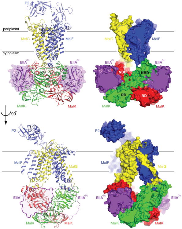
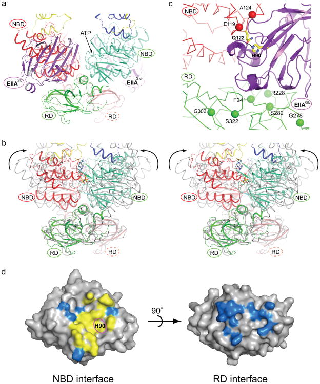
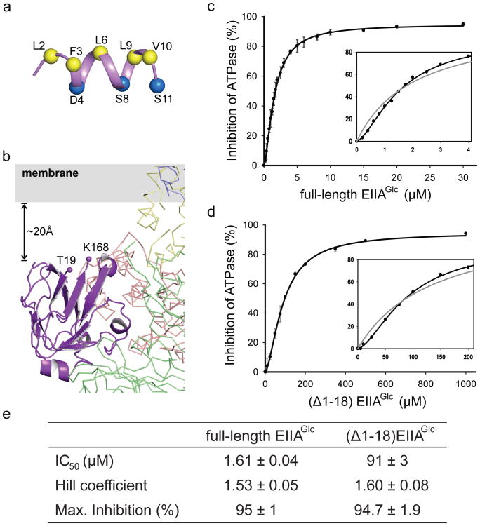
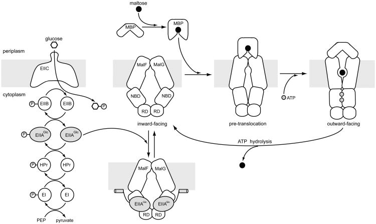
Efficient carbon utilization is critical to the survival of microorganisms in competitive environments. To optimize energy usage, bacteria have developed an integrated control system to preferentially uptake carbohydrates that support rapid growth. The availability of a preferred carbon source, such as glucose, represses the synthesis and activities of proteins necessary for the transport and metabolism of secondary carbon sources. This regulatory phenomenon is defined as carbon catabolite repression. In enteric bacteria, the key player of carbon catabolite repression is a component of the glucose-specific phosphotransferase system, enzyme IIA (EIIA(Glc)). It is known that unphosphorylated EIIA(Glc) binds to and inhibits a variety of transporters when glucose is available. However, understanding the underlying molecular mechanism has been hindered by the complete absence of structures for any EIIA(Glc)-transporter complexes. Here we present the 3.9 Å crystal structure of Escherichia coli EIIA(Glc) in complex with the maltose transporter, an ATP-binding cassette (ABC) transporter. The structure shows that two EIIA(Glc) molecules bind to the cytoplasmic ATPase subunits, stabilizing the transporter in an inward-facing conformation and preventing the structural rearrangements necessary for ATP hydrolysis. We also show that the half-maximal inhibitory concentrations of the full-length EIIA(Glc) and an amino-terminal truncation mutant differ by 60-fold, consistent with the hypothesis that the amino-terminal region, disordered in the crystal structure, functions as a membrane anchor to increase the effective EIIA(Glc) concentration at the membrane. Together these data suggest a model of how the central regulatory protein EIIA(Glc) allosterically inhibits maltose uptake in E. coli.


Figures

Figure 1
Figure 1. Two orthogonal views of the EIIAGlc-MalFGK2 complex
Both a ribbon diagram (left) and a slab view (right) are shown. In the lower panel, EIIAGlc is represented by a transparent surface, with the solid line representing the front molecule and dotted line representing the backside one.
Figure 2
Figure 2. Binding of EIIAGlc prevents MalK closure
a, MalK dimer bound with EIIAGlc. EIIAGlc is shown in purple. The two MalK subunits are coloured in red and green. The nucleotide binding domain (NBD) and regulatory domain (RD) within each MalK subunit are rendered in different shades. The ATP binding site (the Walker A motif) in the front subunit (green) is indicated by an arrow. b, Stereoview of a superposition of the closed MalK dimer, coloured as in panel a, with the open dimer coloured in grey. ATP is shown in stick model. The arrows indicate the rotations of the NBDs necessary to form the closed dimer. c, EIIAGlc-MalK interface. Inducer exclusion resistant mutants are labeled. The active site His 90 of EIIAGlc and Gln 122 of MalK are shown in stick models, and the Hydrogen bond between His 90 and Gln 122 is indicated by a dashed line. d, Surface presentations of EIIAGlc. Residues located within 4.5 Å from the NBD (left) or RD (right) are coloured. Yellow, the 11 residues (Val 39, Val 40, Phe 41, Ile 45, Val 46, Lys 69, Phe 71, Glu 72, Phe 88, His 90, and Val 96) involved in interacting with all four EIIAGlc partners: MalK, glycerol kinase, HPr, and EIIBGlc. The phosphorylation site His 90 is labeled. Blue, MalK interacting residues that are not part of the canonical binding surface
Figure 3
Figure 3. The N-terminus of EIIAGlc likely functions as a membrane anchor
a, Structure of the N-terminal peptide determined by NMR in dihexanoyl phosphatidylglycerol (PDB 1O53). Hydrophobic residues lining one side of the helix are shown in yellow. Hydrophilic residues on the opposite side of the helix are shown in blue. b, Close-up view of EIIAGlc bound to MalK. The two terminal residues visible in the crystal structure (Thr 19 and Lys 168) are labeled. c,d, Inhibition of the MBP-stimulated ATPase activity of reconstituted nanodiscs by full-length (c) and N-terminal truncated EIIAGlc (d). Data points represent the means ± standard deviation of triplicate measurements. Insets show enlargements of the figures fitted to the Hill equation (black line) and to the Michaelis-Menten equation (grey line). e, Summary table of the kinetics.
Figure 4
Figure 4. Inhibition of the maltose transporter by inducer exclusion
Left, Glucose and other PTS substrates are transported and phosphorylated by five coupled reactions, leading to an increased level of unphosphorylated EIIAGlc. Right, the maltose transport cycle. In the resting state, the open MalK dimer coincides with the inward-facing TM domains. Upon association of MBP, the transporter undergoes a conformational change (the pre-translocation state) that permits ATP to promote a concerted motion of MalK closure, reorientation of the TM domains, and opening of MBP. Formation of the outward-facing conformation transfers maltose from MBP to the TM binding site, and at the same time positions ATP at the catalytic site for hydrolysis. ATP hydrolysis releases maltose into the cytoplasm and resets the transporter to the resting state. Under conditions subject to inducer exclusion, unphosphorylated EIIAGlc binds and stabilizes the resting state transporter, thus inhibiting maltose transport.

Similar articles

Cited by

References

    1. Gorke B, Stulke J. Carbon catabolite repression in bacteria: many ways to make the most out of nutrients. Nat Rev Microbiol. 2008;6:613–624. - PubMed
    1. Deutscher J, Francke C, Postma PW. How phosphotransferase system-related protein phosphorylation regulates carbohydrate metabolism in bacteria. Microbiol Mol Biol Rev. 2006;70:939–1031. - PMC - PubMed
    1. Wang G, Peterkofsky A, Clore GM. A novel membrane anchor function for the N-terminal amphipathic sequence of the signal-transducing protein IIAGlucose of the Escherichia coli phosphotransferase system. J Biol Chem. 2000;275:39811–39814. - PubMed
    1. Wang G, Keifer PA, Peterkofsky A. Solution structure of the N-terminal amphitropic domain of Escherichia coli glucose-specific enzyme IIA in membrane-mimetic micelles. Protein Sci. 2003;12:1087–1096. - PMC - PubMed
    1. Hogema BM, et al. Inducer exclusion in Escherichia coli by non-PTS substrates: the role of the PEP to pyruvate ratio in determining the phosphorylation state of enzyme IIAGlc. Mol Microbiol. 1998;30:487–498. - PubMed

Publication types

MeSH terms

Substances

Associated data