. 2013 Jul 18;499(7458):341-5.
doi: 10.1038/nature12348.

Activity-dependent phosphorylation of MeCP2 threonine 308 regulates interaction with NCoR

Affiliations

Activity-dependent phosphorylation of MeCP2 threonine 308 regulates interaction with NCoR

Daniel H Ebert et al. Nature. .

Abstract

Rett syndrome (RTT) is an X-linked human neurodevelopmental disorder with features of autism and severe neurological dysfunction in females. RTT is caused by mutations in methyl-CpG-binding protein 2 (MeCP2), a nuclear protein that, in neurons, regulates transcription, is expressed at high levels similar to that of histones, and binds to methylated cytosines broadly across the genome. By phosphotryptic mapping, we identify three sites (S86, S274 and T308) of activity-dependent MeCP2 phosphorylation. Phosphorylation of these sites is differentially induced by neuronal activity, brain-derived neurotrophic factor, or agents that elevate the intracellular level of 3',5'-cyclic AMP (cAMP), indicating that MeCP2 may function as an epigenetic regulator of gene expression that integrates diverse signals from the environment. Here we show that the phosphorylation of T308 blocks the interaction of the repressor domain of MeCP2 with the nuclear receptor co-repressor (NCoR) complex and suppresses the ability of MeCP2 to repress transcription. In knock-in mice bearing the common human RTT missense mutation R306C, neuronal activity fails to induce MeCP2 T308 phosphorylation, suggesting that the loss of T308 phosphorylation might contribute to RTT. Consistent with this possibility, the mutation of MeCP2 T308A in mice leads to a decrease in the induction of a subset of activity-regulated genes and to RTT-like symptoms. These findings indicate that the activity-dependent phosphorylation of MeCP2 at T308 regulates the interaction of MeCP2 with the NCoR complex, and that RTT in humans may be due, in part, to the loss of activity-dependent MeCP2 T308 phosphorylation and a disruption of the phosphorylation-regulated interaction of MeCP2 with the NCoR complex.


Conflict of interest statement

Author Information: Reprints and permissions information is available at www.nature.com/reprints. The authors declare no competing financial interests.

Figures

Fig. 1
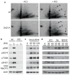
Fig. 1. Phosphotryptic mapping of MeCP2 identifies activity-dependent phosphorylations sites
a, Dissociated cortical cultures, derived from wild-type or MeCP2 S421A KI mice, were metabolically labeled with 32P-orthophosphate and were left untreated or membrane depolarized with KCl. Phospho-peptides were spotted in the lower left corner of the plate, resolved by thin-layer electrophoresis in the horizontal direction and thin-layer chromatography in the vertical direction. The activity-induced spots are indicated (arrows a–c). b, Mice were injected with kainic acid to induce seizures. c, Cortical neurons were stimulated with bicuculline. d, Cortical neurons were membrane depolarized with KCl or treated with BDNF or forskolin. Minutes of stimulation indicated. Lysates were examined by Western blotting with indicated antibodies. Findings confirmed with at least three biological replicates.
Fig. 2
Fig. 2. Phosphorylation of T308 regulates MeCP2’s interaction with the NCoR complex
a, Peptides corresponding to MeCP2, either not phosphorylated (np) or synthesized to be phosphorylated at the amino acid residue corresponding to 308 (p), were incubated with cortical neuron lysates. Pull-downs were visualized by Western blotting with indicated antibodies. b, Peptide pull-downs using synthetic peptides (at 10x or 1X concentrations) containing one of the following mutations, R306C, T308A, T308D, or T308E. c, Luciferase reporter gene assay with plasmids encoding reporter and GAL4-MeCP2 variants, as indicated, transfected into cortical neurons. “None” had no GAL4-MeCP2 variant transfected. Shown are averages and s.e.m. of three biological replicates. Findings for each experiment were confirmed with at least three biological replicates.
Fig. 3
Fig. 3. MeCP2 T308A KI mice have altered activity-dependent gene expression
a, Dissociated cortical neuron cultures, generated from MeCP2 T308A KI mice (KI) and wild-type littermates (wt) were membrane depolarized with KCl. Shown are fold-inductions of mRNA levels measured by RT-PCR for the average of three independent days of dissection (biological replicates). For Npas4, p-values were 0.024 (repeated measures ANOVA) and 0.03 at 1 h and 0.01 at 6 h (paired two-tailed t-test). b, Eight-week-old mice, kept in the dark for previous two weeks, were either kept in the dark (0 h) or exposed to a light source for six hours (6 h). From dissected visual cortices, RT-PCR values were divided by the average of wt 6 h. The number of littermate mice averaged per timepoint were: wt 0 h (n=6), KI 0 h (n=6), wt 6 h (n=8), and KI 6 h (n=10). Error bars indicate s.e.m. For Npas4 at 6 h, the two-tailed t-test p-value is 0.0007 and for Bdnf at 6 h, p-value is 0.04.
Fig. 4
Fig. 4. Phosphorylation of MeCP2 T308 is a key mechanism underlying RTT
a, MeCP2 R306C KI mice and wild-type littermates (wt) were treated with kainic acid (KA) to induce seizures or left untreated (-). Forebrain lysates were resolved by Western blot analysis with indicated antibodies. Findings confirmed with five biological replicates. b, Brains were dissected and weighed for T308A KI mice (n=16) and wild-type littermates (n=13) with 5% reduction in the KI and p-value between genotypes of 0.00006 (unpaired two-tailed t-test). c, T308A KI mice (n=16) and wild-type littermates (n=13) were scored for the presence of a hindlimb clasp. P-value was 0.00005 (two-proportion Z-test). d, T308A KI mice (n=16) and wild-type littermates (n=13) were placed on an accelerating rotarod, and latency to fall was measured with a p-value between genotypes of 0.02 (two-tailed t-test). e, T308A KI mice (n=17) and wild-type littermates (n=15) were injected with 40 mg/kg PTZ and latency to onset of generalized tonic-clonic seizures was measured. P-value was 0.029 (two-sample Kolmogorov-Smirnov test). Error bars indicate s.e.m.

References

    1. Nan X, Campoy FJ, Bird A. Mecp2 Is a Transcriptional Repressor with Abundant Binding Sites in Genomic Chromatin. Cell. 1997;88:471–481. - PubMed
    1. Nan X, et al. Transcriptional Repression by the Methyl-Cpg-Binding Protein Mecp2 Involves a Histone Deacetylase Complex. Nature. 1998;393:386–389. - PubMed
    1. Amir RE, et al. Rett Syndrome Is Caused by Mutations in X-Linked Mecp2, Encoding Methyl-Cpg-Binding Protein 2. Nat Genet. 1999;23:185–188. - PubMed
    1. Chahrour M, et al. Mecp2, a Key Contributor to Neurological Disease, Activates and Represses Transcription. Science. 2008;320:1224–1229. - PMC - PubMed
    1. Skene PJ, et al. Neuronal Mecp2 Is Expressed at near Histone-Octamer Levels and Globally Alters the Chromatin State. Mol Cell. 2010;37:457–468. - PMC - PubMed

Publication types