. 2013 Aug;148(2):177-85.
doi: 10.1016/j.clim.2013.05.008. Epub 2013 May 22.

Immunoglobulin G1 and immunoglobulin G4 antibodies in multiple sclerosis patients treated with IFNβ interact with the endogenous cytokine and activate complement

Affiliations

Immunoglobulin G1 and immunoglobulin G4 antibodies in multiple sclerosis patients treated with IFNβ interact with the endogenous cytokine and activate complement

Swaminathan Sethu et al. Clin Immunol. 2013 Aug.

Abstract

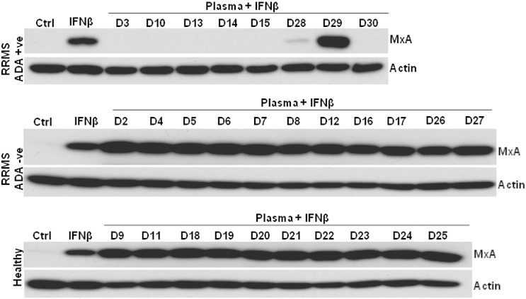
A subset of patients with relapsing-remitting multiple sclerosis (RRMS) on therapy with interferon beta (IFNβ) develop neutralising anti-drug antibodies (ADA) resulting in reduced, or loss of, therapeutic efficacy. The aims were to characterise the relative contributions of anti-IFNβ antibody isotypes to drug neutralising activity, ability of these antibodies to cross-react with endogenous IFNβ, to form immune complexes and activate complement. IFNβ-specific ADA were measured in plasma from RRMS patients treated with IFNβ1a (Rebif(®)). Neutralisation of endogenous and therapeutic IFNβ by ADA was determined by IFNβ bioassay. IFNβ-ADA profile was predominantly comprised of IgG1 and IgG4 antibody isotypes. The contribution of IgG4-ADA towards neutralising activity was found to be minimal. Neutralising IFNβ-ADA blocks endogenous IFNβ activity. ADA interaction with therapeutic IFNβ results in immune complex formation and complement activation. In summary, IgG1 and IgG4 IFNβ-ADA have the ability to neutralise therapeutic and endogenous protein and to activate complement.

Keywords: Anti-drug antibody;; Complement; Immunogenicity;; Interferon beta;; Neutralising antibody;; Relapsing-remitting multiple sclerosis;.


Figures

Figure 1
Figure 1
Immunoglobulin types/subtypes of therapeutic IFNβ-ADA. Total and IFNβ-specific IgG (a and c) and IgM (b and d) in the plasma of healthy donors (healthy) and RRMS patients (RRMS) were determined by ELISA. Data points ▼ in graph (a) are categorised as IFNβ-specific IgG based ADA positive samples. (e) Total IgG subclass in healthy and RRMS plasma samples. (f) IFNβ-specific IgG subclass levels in 8 ADA positive samples detected. (g) Proportion of IFNβ-specific IgG subclass in each ADA positive plasma sample. Statistical analyses include t-test and ANOVA. p = 0.0352 (t-test unpaired); ⁎⁎p = 0.0045 (ANOVA); healthy — n = 10; RRMS — n = 19. The data point in each graph is a mean of triplicate measurements and is a representative of three independent experiments.
Figure 2
Figure 2
Neutralisation potential of therapeutic IFNβ-ADA. Immunoblot shows IFNβ-induced MxA protein levels in THP-1 cells. Therapeutic IFNβ (100U Rebif) pre-incubated with 1:10 dilution of plasma samples of healthy and RRMS with (ADA + ve) and without ADA (ADA − ve) for 1 hour at 37 °C was added to THP-1 cells for 18 hours. Proteins from whole cell lysates of these cells were separated by SDS–PAGE and western immunoblotting performed. Membranes were probed for human MxA and β-actin. “Ctrl” indicates cell lysates prepared from cells without any treatment and “IFNβ” indicates lysates from cells treated with 100U Rebif only. D2–D30 are lysate samples from cells incubated with the plasma and IFNβ mixture. The images are representative of three independent experiments.
Figure 3
Figure 3
Neutralisation potential of IgG4 based therapeutic IFNβ-ADA. (a) “Neutralisation (%)” is the percentage based obtained on densitometry analysis immunoblot as shown in Fig. 3b assessing IFNβ-induced MxA neutralisation by RRMS ADA + ve plasma samples with and without IgG4 relative to “IFNβ” only category. The percentage indicates the difference in the neutralisation potential of IgG4 depleted plasma compared to its respective non-depleted fraction. (b) Immunoblot exhibits variable differences in the potency of ADA positive plasma samples with and without IFNβ-specific IgG4 to neutralise IFNβ-induced MxA protein levels in THP-1 cells. Therapeutic IFNβ (100U Rebif) pre-incubated with diluted of plasma from 8 RRMS ADA + ve samples (1:1000 — D3, D10, D13, D14, D15 and 1:10 — D28, D30) with (Pre) and without (Post) IgG4 for 1 hour at 37 °C was added to THP-1 cells for 18 hours. Proteins from whole cell lysates of these cells for each category were electrophoretically (SDS–PAGE) separated and probed for human MxA. β-Actin probed for all the samples served as loading controls. “Ctrl” indicates cell lysates prepared from cells without any treatment and “IFNβ” indicate lysates from cells treated with 100U Rebif only. The images are representatives of three independent experiments.
Figure 4
Figure 4
IFNβ-specific ADA cross reacts and neutralises endogenous IFNβ-induced bioactivity. (a) Bioactivity of endogenous IFNβ was assessed by triggering IFNβ induced MxA by TLR4/3 (LPS — 1 μg/ml)/(poly(I:C) — 10 μg/ml) activation. Cross reactivity of ADA to endogenous IFNβ was studied by investigating whether ADA were able to block or inhibit the induction of MxA in this system. (b) Immunoblot image shows MxA protein levels induced by LPS and poly(I:C) (LPS + PIC) stimulation for 24 hours in THP-1 cells. Induction of MxA is as a result of endogenous Type 1 interferons, including IFNβ triggered by LPS + PIC. The immunoblot also indicates the changes in the level of induced MxA when the cells were co-incubated with 1:10 dilution of plasma samples from 8 RRMS ADA + ve (D3, D10, D13, D14, D15, D28, D29 and D30) individuals along with LPS + PIC. Proteins from whole cell lysates for each category were electrophoretically (SDS–PAGE) separated and probed for human MxA. β-Actin probed for all the samples served as loading controls. “Ctrl” indicates cell lysates prepared from cells without any treatment and “LPS + PIC” indicates lysates from cells treated with LPS + PIC only. “Neutralisation (%)” is the percentage (densitometric analysis) of LPS + PIC induced MxA neutralised by RRMS ADA + ve plasma samples relative to “LPS + PIC” only category. Immunoblot shown here is a representative of three independent experiments.
Figure 5
Figure 5
Therapeutic ADA-IFNβ immune complex activates complement system. (a) Ag[IFNβ]:Ab[ADA] ratio (IFNβ:ADA) at which immune complexes (IC) were formed by incubating IFNβ with serum containing ADA for 1 hour at 37 °C (n = 2). Graphs indicate the levels of ADA-IFNβ immune complexes bound to C1q (b and c) and subsequent changes in the level of C3a (d and e) in 8 RRMS ADA + ve (D3, D10, D13, D14, D15, D28, D29 and D30) serum samples. Statistical analyses include t-test and ANOVA. p = 0.0180 (t-test paired); ⁎⁎p < 0.0001 (t-test paired); #p < 0.0001 (ANOVA); n = 8. The data point in each graph is a mean with standard deviation of triplicate measurements and is a representative of three independent experiments.

References

    1. Compston A., Coles A. Multiple sclerosis. Lancet. 2008;372:1502–1517. - PubMed
    1. Li D.K., Paty D.W. Magnetic resonance imaging results of the PRISMS trial: a randomized, double-blind, placebo-controlled study of interferon-beta1a in relapsing-remitting multiple sclerosis. Prevention of Relapses and Disability by Interferon-beta1a Subcutaneously in Multiple Sclerosis. Ann. Neurol. 1999;46:197–206. - PubMed
    1. Paty D.W., Li D.K. Interferon beta-1b is effective in relapsing-remitting multiple sclerosis. II. MRI analysis results of a multicenter, randomized, double-blind, placebo-controlled trial. UBC MS/MRI Study Group and the IFNB Multiple Sclerosis Study Group. Neurology. 1993;43:662–667. - PubMed
    1. Perini P., Calabrese M., Biasi G., Gallo P. The clinical impact of interferon beta antibodies in relapsing-remitting MS. J. Neurol. 2004;251:305–309. - PubMed
    1. Sorensen P.S., Koch-Henriksen N., Ross C., Clemmesen K.M., Bendtzen K. Appearance and disappearance of neutralizing antibodies during interferon-beta therapy. Neurology. 2005;65:33–39. - PubMed

Publication types