. 2013 Aug;14(8):831-9.
doi: 10.1038/ni.2622. Epub 2013 Jun 16.

The nuclear receptor LXRα controls the functional specialization of splenic macrophages

Affiliations

The nuclear receptor LXRα controls the functional specialization of splenic macrophages

Noelia A-Gonzalez et al. Nat Immunol. 2013 Aug.

Abstract

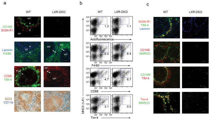
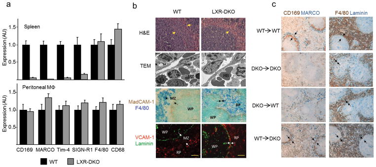
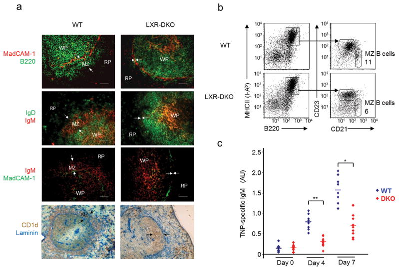
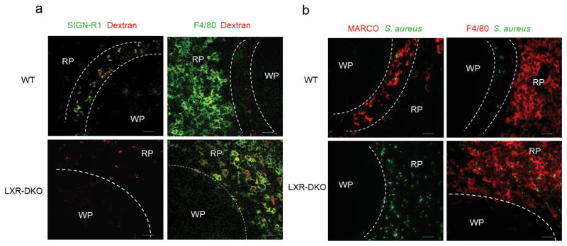
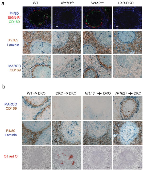
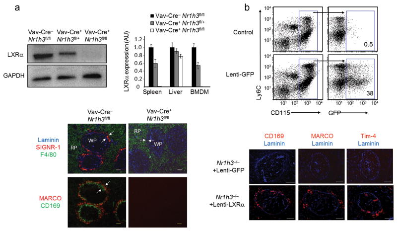
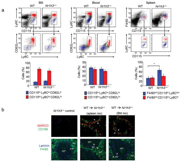
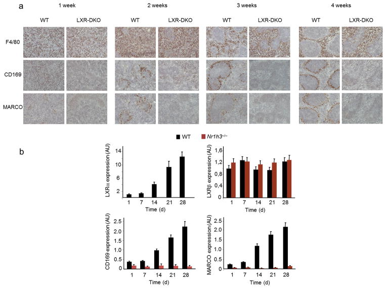
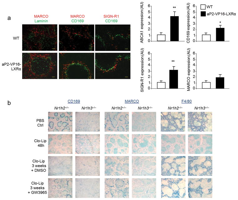
Macrophages are professional phagocytic cells that orchestrate innate immune responses and have considerable phenotypic diversity at different anatomical locations. However, the mechanisms that control the heterogeneity of tissue macrophages are not well characterized. Here we found that the nuclear receptor LXRα was essential for the differentiation of macrophages in the marginal zone (MZ) of the spleen. LXR-deficient mice were defective in the generation of MZ and metallophilic macrophages, which resulted in abnormal responses to blood-borne antigens. Myeloid-specific expression of LXRα or adoptive transfer of wild-type monocytes restored the MZ microenvironment in LXRα-deficient mice. Our results demonstrate that signaling via LXRα in myeloid cells is crucial for the generation of splenic MZ macrophages and identify an unprecedented role for a nuclear receptor in the generation of specialized macrophage subsets.


Conflict of interest statement

Competing financial interests

The authors declare no competing financial interests

Figures

Figure 1
Figure 1. Defective MZ macrophage differentiation in LXR-DKO mice
(a) Top, splenic sections from wild-type (WT) and LXR-DKO mice were stained with specific antibodies that detect CD169+ macrophages (green) and SIGN-R1+ macrophages (red). Below, consecutive sections show RP macrophages (F4/80, green) and stromal endothelial cells (laminin, blue): CD68+ macrophages (red) and TIM-4+ cells in the MZ (green). Bottom, immunohistochemical analysis of RP CD11b+ cells (blue) and B cells (B220, brown). Arrows indicate the RP and WP borders. Scale bars represent 50 μm. (b) Flow cytometry analysis of F4/80, CD68, Tim-4 and MHCII expression in splenic cell suspensions from WT and LXR-DKO mice. Representative plots are shown from three independent experiments with n = 4–5 mice per genotype. Numbers within plots indicate percent of gated cell population (c) Immunofluorescence analysis of spleen sections from WT and LXR-DKO mice showing combinations of double or triple stainings with antibodies that recognize MZ macrophages (SIGN-R1, MARCO and Tim-4), marginal metallophillic macrophages (CD169) or endothelial cells (Laminin). Scale bars represent 50 μm. Representative images obtained from five independent experiments with n= 3–5 mice per genotype (a, c).
Fig. 2
Fig. 2. Generation of MZ macrophages require LXR function in hematopoietic progenitors
(a) Relative mRNA abundance of SIGN-R1, MARCO, Tim-4, F4/80 and CD68 in WT and LXR-DKO spleens (top) and peritoneal macrophages (bottom). Data are normalized to ribosomal protein Rplp0 (also called 36B4) mRNA expression. Bars indicate mean +/− SEM (n=3 mice). Data are representative of two independent experiments. (b) Hematoxilin and eosin staining (top), transmission electron microscopy images (second row); abbreviations: MZ: MZ macrophage; m: marginal metallophilic macrophage; F: follicular reticular cell; RB: red blood cell. RP: RP macrophage. Third row shows immunohistochemical localization of RP F4/80+ macrophages relative to MadCAM-1 marginal sinus cells; (bottom row) VCAM-1+ cells in the RP relative to laminin+ endothelial cells; scale bar represents 30 αm. Arrows indicate RP and WP borders. Representative images obtained from three independent experiments with n = 2–3 mice per group. (c) Immunohistochemical localization of RP and MZ compartments in WT and LXR-DKO mice after reciprocal bone marrow transplants. Scale bar represents 50 αm. Arrows indicate the RP and WP borders. Marginal metallophilic macrophages (CD169, brown) and MZ macrophages (MARCO, blue), or RP macrophages (F4/80, brown) and endothelial cells (Laminin, blue) were stained in consecutive spleen sections obtained from mice 12 weeks after BM transplantation (representative images obtained from two independent experiments with n = 5–6 mice per group).
Fig. 3
Fig. 3. Analysis of MZ B cell distribution and function
(a) Immunofluorescence and immunohistochemical analysis of spleen sections from WT and LXR-DKO mice stained with antibodies that detect MZ B cells. Top, MZ B220+ B cells identified outside the follicles, which are delimitated by MadCAM-1+ cells in WT mice and absent in LXR-DKO mice; second row IgMhiIgDlo MZ B cells; third row IgMhi MZ B cells outside MadCAM-1+ sinus lining cells; and bottom CD1d Laminin double immunohistochemical analysis. Arrows indicate the RP and WP borders. Scale bars represent 50 μm. Representative images from n=10 mice per genotype are shown (b) Flow cytometry of splenocytes from WT and LXR-DKO mice stained with CD23 and CD21 antibodies and analyzed in gated B220+ cells. MZ B cells were quantifed as CD21hiCD23lo; representative analysis obtained from three independent experiments of n = 3–5 mice is presented. (c) Mice were immunized i.v. with TNP-ficoll and serum samples were obtained before immunization and 4 and 7 days after immunization. TNP-specific IgM titers were measured by ELISA. Representative results obtained from two independent experiments with n = 6–10 mice per group are shown. Each (◆) symbol represents individual WT (blue, n = 8) or LXR-DKO (red, n = 9) mice; *P < 0.01 and **P < 0.001.
Fig. 4
Fig. 4. Abnormal capture of blood-borne antigens by LXR-DKO splenic macrophages
Immunofluorescence microscopy of spleen sections obtained from WT and LXR-DKO mice after 30 min post i.v. injection with dextran-Alexa Fluor 594 (a) and Alexa Fluor 488-Staphyloccocus aureus particles (b). MZ macrophages were detected with SIGN-R1 (green) and MARCO (red) abs in (a) and (b) respectively. RP macrophages were detected with F4/80 antibody (green in a; red in b). Dashed lines delimitate the border of the WP and the RP. Scale bar represents 50 μm. Representative pictures from four independent experiments with n = 3–5 mice per group are presented.
Fig. 5
Fig. 5. MZ macrophage differentiation is specifically controlled by LXRα
(a) Top, immunofluorescence analysis of spleen sections from WT, LXRα-KO, LXRβ-KO, and LXR-DKO mice stained with CD169, SIGN-R1 and F4/80 antibodies. Middle, Immunohistochemical analysis of F4/80+ RP macrophages (brown) relative to Laminin+ endothelial cells (blue); bottom, consecutive sections stained for MARCO+ (blue) and CD169+ cells (brown). (b) Consecutive spleen sections (top, middle) show immunohistochemical localization of MZ macrophages (MARCO, blue and CD169, brown), and F4/80+ (brown) and Laminin+ endothelial cells (blue) obtained from LXR-DKO mice after transplant experiments with bone marrow from the indicated donor mice. Bottom, Oil-Red O staining in spleen sections from the indicated mice after bone marrow transplantation. Scale bar represents 50 μm. Representative pictures from four independent experiments with n = 3–5 mice (panel a) and two independent experiments with n= 6 mice per group (panel b) are presented.
Fig. 6
Fig. 6. LXRα function in myeloid cells is required for MZ macrophage development
(a) Immunoblot analysis of LXRα and GADPH in peritoneal macrophages from Vav-CreNr1h3fl/fl, Vav-Cre+Nr1h3fl/+, and Vav-Cre+Nr1h3fl/fl mice. Right, relative mRNA abundunce of LXRα in spleen, liver and bone marrow derived macrophages (BMDM) from Vav-CreNr1h3fl/fl, Vav-Cre+Nr1h3fl/+, and Vav-Cre+Nr1h3fl/fl mice. Data are normalized to 36B4 mRNA expression. Data represent mean +/− SEM (n= 2–3 mice). Data are representative of four independent experiments. Immunofluorescence analysis of consecutive sections obtained from Vav-Cre/Nr1h3fl/fl and Vav-Cre+/Nr1h3fl/fl spleens that were stained with Laminin, SIGN-R1 and F4/80 antibodies (top); bottom, MZ macrophages (MARCO, red), and marginal metallophilic macrophages (CD169, green); scale bar represents 50 μm. Representative images obtained from four independent experiments with n = 3–6 mice per group (b) Gain-of-function studies with a macrophage specific lentivirus expressing LXRα. Top, flow cytometry analysis of blood monocytes from WT mice transplanted with HSCs transduced with Lenti-GFP viral particles. Middle, immunofluorescence analysis of macrophage markers in spleen sections from LXRα-KO mice that were transplanted with LXRα-KO HSCs transduced with Lenti-GFP or Lenti-LXRα particles (representative images from two independent experiments with n = 4–5 mice per group); scale bar represents 100 μm.
Fig. 7
Fig. 7. Adoptive transfer of monocytes reconstitute splenic MZ microenvironment in LXRα-KO mice
(a) Flow cytometry analysis of monocyte subsets in BM, blood and spleen cells from WT and LXRα-KO mice. Classical and non-classical monocytes were detected based on the expression of Ly6C and CD62L in CD115+ cells in BM and blood, and based on the expression of Ly6C in CD115+ F4/80+ cells in spleen. Bottom, percentages of classical and non-classical monocytes in bone marrow, blood and spleen measured by flow cytometry. Bars indicate mean +/− (SEM) from n = 7 mice. Data are representative from four independent experiments. (b) Immunofluorescence analysis of consecutive spleen sections from non-irradiated LXRα-KO mice reconstituted with the indicated WT monocytes from BM or spleen origin. Arrows indicate the RP and WP borders. Representative images from three independent experiments with n = 5–6 mice/group are presented. Scale bar represents 50 μm..
Fig. 8
Fig. 8. LXR signaling is necessary for postnatal development of splenic MZ macrophages
a) Immunohistochemical analysis of marginal metallophilic macrophages (CD169), MZ macrophages (MARCO), and red pulp macrophages (F4/80) in spleen sections obtained from WT and LXR-DKO at the indicated times after birth; representative images from three independent experiments with n= 3–4 mice per genotype are presented; scale bar represents 50 μm. (b) mRNA relative expression of LXRα, LXRβ, CD169, MARCO, in WT and LXRα-KO spleens at the indicated times after birth. Data represents mean +/− (SEM) obtained from 2 independent experiments with n = 6 mice per genotype. Data are normalized to 36B4 mRNA expression.
Fig. 9
Fig. 9. Activation of LXRα accelerates the development and renewal of MZ macrophages
(a) Immunofluorescence analysis of spleen sections from WT and aP2-VP16-LXRα mice at three weeks of age stained with MARCO, SIGN-R1 (MZ macrophages), CD169 (marginal metallophilic macrophages) and Laminin (endothelial cells) antibodies (left); scale bar represents 25 μm. Right, relative mRNA abundance of ABCA1, CD169, SIGN-R1 and MARCO from spleen samples (n = 5) of WT and AP2-VP16-Nr1h3 spleen at three weeks of age. Data obtained from two independent experiments with n = 3–5 mice (b) Immunohistochemistry analysis of CD169, MARCO and F4/80 expression in spleen sections from LXRβ-KO and LXRα-KO mice after clodronate liposome macrophage depletion and GW3965 or vehicle control treatment at the indicated times; scale bar represents 150 μm. Representative images from 2 independent experiments with n = 5–6 mice per group are presented.

References

    1. Taylor PR, et al. Macrophage receptors and immune recognition. Annu Rev Immunol. 2005;23:901–944. - PubMed
    1. Medzhitov R. Origin and physiological roles of inflammation. Nature. 2008;454:428–435. - PubMed
    1. Elliott MR, Ravichandran KS. Clearance of apoptotic cells: implications in health and disease. J Cell Biol. 2010;189:1059–1070. - PMC - PubMed
    1. Gordon S, Taylor PR. Monocyte and macrophage heterogeneity. Nat Rev Immunol. 2005;5:953–964. - PubMed
    1. Hashimoto D, Miller J, Merad M. Dendritic cell and macrophage heterogeneity in vivo. Immunity. 2011;35:323–335. - PMC - PubMed

Publication types

MeSH terms