. 2013 Aug 2;288(31):22621-35.
doi: 10.1074/jbc.M113.481382. Epub 2013 Jun 13.

Serotype-specific differences in dengue virus non-structural protein 5 nuclear localization

Affiliations

Serotype-specific differences in dengue virus non-structural protein 5 nuclear localization

Holger Hannemann et al. J Biol Chem. .

Abstract

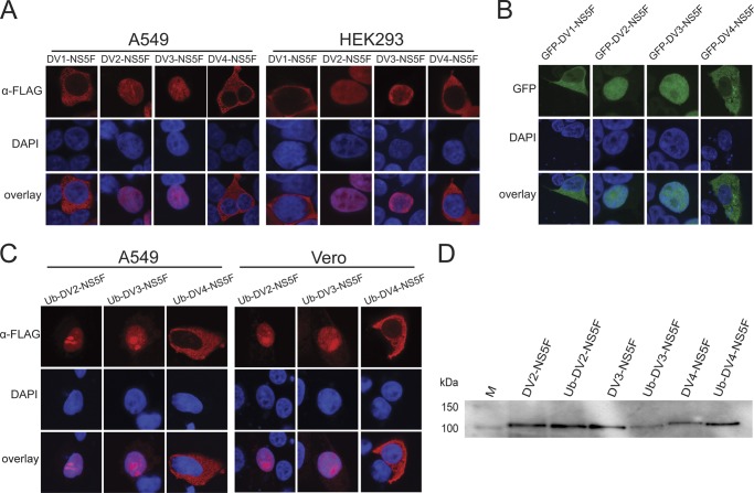
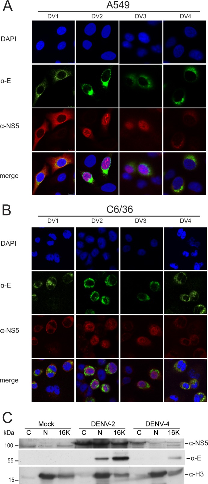
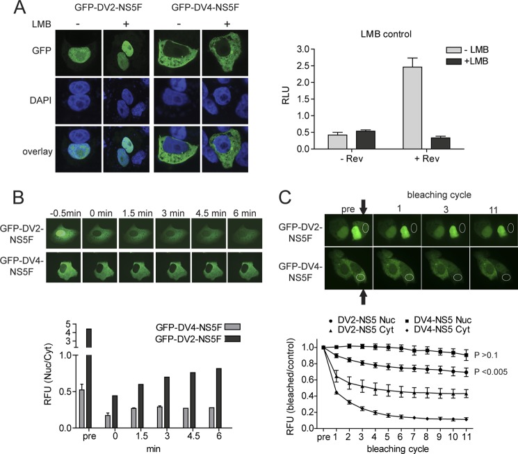
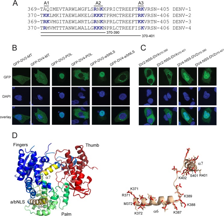
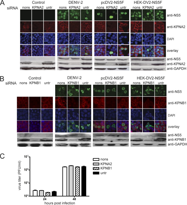
The four serotypes of dengue virus (DENV-1 to -4) cause the most important arthropod-borne viral disease of humans. DENV non-structural protein 5 (NS5) contains enzymatic activities required for capping and replication of the viral RNA genome that occurs in the host cytoplasm. However, previous studies have shown that DENV-2 NS5 accumulates in the nucleus during infection. In this study, we examined the nuclear localization of NS5 for all four DENV serotypes. We demonstrate for the first time that there are serotypic differences in NS5 nuclear localization. Whereas the DENV-2 and -3 proteins accumulate in the nucleus, DENV-1 and -4 NS5 are predominantly if not exclusively localized to the cytoplasm. Comparative studies on the DENV-2 and -4 NS5 proteins revealed that the difference in DENV-4 NS5 nuclear localization was not due to rapid nuclear export but rather the lack of a functional nuclear localization sequence. Interaction studies using DENV-2 and -4 NS5 and human importin-α isoforms failed to identify an interaction that supported the differential nuclear localization of NS5. siRNA knockdown of the human importin-α isoform KPNA2, corresponding to the murine importin-α isoform previously shown to bind to DENV-2 NS5, did not substantially affect DENV-2 NS5 nuclear localization, whereas knockdown of importin-β did. The serotypic differences in NS5 nuclear localization did not correlate with differences in IL-8 gene expression. The results show that NS5 nuclear localization is not strictly required for virus replication but is more likely to have an auxiliary function in the life cycle of specific DENV serotypes.

Keywords: Dengue; Flaviviruses; Nuclear Transport; Positive Strand RNA Viruses; Viral Polymerase; Virology.


Figures

FIGURE 1.
FIGURE 1.
Subcellular localization of transiently expressed DENV-1 to -4 NS5. A, CLSM images of A549 (left panels) or HEK293 cells (right panels) transfected with pcDV1-NS5F (DV1-NS5F), pcDV2-NS5F (DV2-NS5F), pcDV3-NS5F (DV3-NS5F), and pcDV4-NS5F (DV4-NS5F) and immunostained using an anti-FLAG antibody (α-FLAG) at 24 h post-transfection. B, CLSM images of A549 cells transfected with pcGFP-DV1-NS5F (GFP-DV1-NS5F), pcGFP-DV2-NS5F (GFP-DV2-NS5F), pcGFP-DV3-NS5F (GFP-DV3-NS5F), and pcGFP-DV4-NS5F (GFP-DV4-NS5F) and analyzed for GFP fluorescence at 24 h post-transfection. C, CLSM images of A549 (left panels) and Vero cells (right panels) transfected with pcUb-DV2-NS5F (Ub-DV2-NS5F), pcUb-DV3-NS5F (Ub-DV3-NS5F), and pcUb-DV4-NS5F (Ub-DV4-NS5F) and immunostained using an anti-FLAG antibody at 24 h post-transfection. Nuclear DNA was stained with DAPI in all experiments. D, HEK293 cells were transfected with the plasmids described in A and C. At 24 h post-transfection, total cell lysates were prepared and analyzed by SDS-PAGE and Western blotting. The respective NS5 proteins were detected using an anti-FLAG antibody. The positions of relevant molecular mass markers (M) are shown.
FIGURE 2.
FIGURE 2.
Subcellular localization of DENV-1 to -4 NS5 during viral infection. CLSM images of A549 cells (A) and insect C6/36 cells (B) infected with DENV-1, -2, -3 and -4 at a multiplicity of infection of 2.0 and immunostained using anti-E (α-E) and anti-NS5 (α-NS5) antibodies at 30 h postinfection. In each panel, a selection of infected (as evidenced by anti-E staining) and non-infected cells are shown as a control for nonspecific staining by the anti-NS5 antibody. Nuclear DNA was stained with DAPI. C, Western blot analysis of cytoplasmic (C), nuclear (N), and cytoplasmic membrane (16K) fractions prepared from Huh-7 cells at 30 h postinfection with DENV-2 or -4 (multiplicity of infection of 3) or mock-infected. 10 μg of protein from each cellular fraction was analyzed for the presence of the DENV NS5 and E proteins and histone H3 using specific antibodies by Western blotting. Histone H3 was used as a nuclear marker. The positions of relevant molecular mass markers are shown in kDa.
FIGURE 3.
FIGURE 3.
Analysis of DENV-2 and -4 NS5 nuclear-cytoplasmic trafficking. A, Vero cells were transfected with either pcGFP-DV2-NS5F (GFP-DV2-NS5F) or pcGFP-DV4-NS5F (GFP-DV4-NS5F). At 16 h post-transfection, the cells were treated with either 15 μg/ml LMB (+) or DMSO (−). 6 h post-treatment, GFP fluorescence was analyzed by CLSM. Nuclear DNA was stained with DAPI. The inhibition of CRM1 by LMB was confirmed using a CRM1-dependant luciferase-based reporter system. Vero cells were transfected with either pLUCSALRRE, which expresses a transcript containing an HIV rev-responsive element and encodes luciferase (−Rev), or pLUCSALRRE and a plasmid encoding HIV rev (+Rev). The cells were treated with 15 μg/ml LMB after transfection. At 16 h post-transfection, the luciferase activity in the cells was measured and expressed as relative light units (RLU). Error bars, measurement averages of three independent experiments. B, Vero cells were transfected with either pcGFP-DV2-NS5F (GFP-DV2-NS5F) or pcGFP-DV4-NS5F (GFP-DV4-NS5F). At 24 h post-transfection, the movement of NS5 between the cytoplasm and nucleus of transfected cells was analyzed by FRAP. The nuclei of transfected cells (n = 5) were bleached, and the relative ratios of nuclear versus cytoplasmic fluorescence (RFU (Nuc/Cyt)) at the given times postbleaching were determined. Representative images of the photobleaching of one transfected cell are shown. The images of cells expressing GFP-DV2-NS5F were purposely overexposed to visualize the cytoplasmic fraction of NS5, whereas the original pictures were used for quantification. C, Vero cells were transfected as in B. For FRAP analysis, the indicated region (white circle) of the cytoplasm of a transfected cell was bleached, and CLSM images were taken 29 s later. This sequence was repeated 11 times. Representative images are shown prebleaching and for the first, third, and eleventh bleaching cycles. A bleached cell (arrow) is shown beside an untreated cell for comparison. The images were used to determine the relative fluorescence (RFU) intensities of the cytoplasm (triangles and diamonds) or nuclei (squares and circles) in bleached versus unbleached cells (n = 5; error bars, S.D.), with all prebleached RFU values set to 1. The significance of the variation between the nuclear fluorescence before bleaching and after 11 bleaching cycles was determined using a two-tailed Student's t test and is shown for cells expressing GFP-DV2-NS5F and GFP-DV4-NS5F.
FIGURE 4.
FIGURE 4.
DENV-4 NS5 does not have a functional a/bNLS. A, alignment of the DENV-1, -2, -3, and 4 NS5 amino acid sequences corresponding to the DENV-2 NS5 a/bNLS. Three basic amino acid clusters found in the DENV-2 a/bNLS (termed A1, A2, and A3) and conserved basic amino acid sequences in the other sequences are shown in boldface type and colored blue. The amino acid sequences interchanged between DENV-2 and -4 NS5 are shown. B, CLSM images of A549 cells transfected with pcGFP-DV2-MT (GFP-DV2-MT) and pcGFP-DV4-MT (GFP-DV4-MT) (left panels), pcGFP-DV2-POL (GFP-DV2-POL) and pcGFP-DV4-POL (GFP-DV4-POL) (middle panels), and pc2xGFP-DV2-NLS (GFP-DV2-a/bNLS) and pc2xGFP-DV4-NLS (GFP-DV4-a/bNLS) (right panels). C, CLSM images of A549 cells transfected with pcGFP-DV2-NS5-DV4(370–390) (DV2-NS5-DV4370–390), pcGFP-DV2-NS5-DV4(370–401) (DV2-NS5-DV4370–401), pcGFP-DV4-NS5-DV2(370–390) (DV4-NS5-DV2370–390), and pcGFP-DV4-NS5(370–401) (DV4-NS5-DV2370–401). The transfected cells were analyzed for GFP fluorescence at 24 h post-transfection. Nuclear DNA was stained with DAPI in all experiments. D, superimposition of models of the POL structures of DENV-2 NS5 and DENV-2 NS5 containing DENV-4 amino acids 370–401. The DENV-2 POL sequences were used to generate hidden Markov models (72), which were then compared against a database of hidden Markov models with solved atomic structures. The MODELLER software suite (73) was used to generate the three-dimensional models based upon the x-ray structure of the DENV-3 NS5 POL domain (60). PyMOL (74) was the used to align the structures and for molecular graphics. The aligned POL structures are shown on the left, with the fingers, thumb, and palm subdomains and the a/bNLS (containing α helices 6 and 7) shaded in blue/light blue, red/salmon, green/pale green, and yellow/sand for the DENV-2 and DENV-2/4 POLs, respectively. On the right, an expanded view of the a/bNLS alignment is shown in schematic form. Amino acids that differ in side chain residue or orientation are shown in stick form. The amino acids in the A1, A2, and A3 clusters are numbered for the DENV-2 and -4 sequences.
FIGURE 5.
FIGURE 5.
Co-IP analysis of DENV-2 and -4 NS5 interactions. A, HEK293 cells were co-transfected with either a plasmid coding for KPNA2-V5 or no plasmid (con) together with pcDV2-NS5F (DV2-NS5F), pcDV4-NS5F (DV4-NS5F), or pc3xFLAG-Rac1 (Rac1-F). Co-IP analysis was done using anti-V5 beads. The immunoprecipitates (IP) and total cell lysates (Input) were analyzed by SDS-PAGE and Western blotting using an anti-FLAG antibody to detect NS5F and Rac1-F and an anti-V5 antibody to detect KPNA2-V5. *, nonspecific band recognized by the anti-V5 antibody. B, HEK293 cells were co-transfected with either pGFP-DV2-NS5F (GFP-DV2-NS5F), pGFP-DV4-NS5F (GFP-DV4-NS5F), or pEGFP (GFP) together with a plasmid coding for human STAT2. Co-IP analysis was conducted using GFP-Trap beads and TBS containing 150 mm NaCl and 1% Triton X-100 for cell lysis and washing. The immunoprecipitates (IP) and total cell lysates (Input) were analyzed by SDS-PAGE and Western blotting using anti-STAT2 and anti-GFP antibodies to detect STAT2 and GFP-NS5/GFP, respectively. All co-IP assays were done 24 h post-transfection.
FIGURE 6.
FIGURE 6.
Effects of siRNA knockdown of KPNA2 and KPNB1 on NS5 nuclear localization. A, HEK293 cells or HEK293 cells stably expressing DENV-2 NS5 (HEK-DV2-NS5F), in response to doxycycline induction, were transfected with either a non-sense (nons) or a KPNA2 siRNA pool or left untreated (untr). 72 h post-transfection, HEK293 cells were either not treated (Control), infected with DENV-2, or transfected with pcDV2-NS5F. The HEK-DV2-NS5F cells were treated with doxycycline to induce NS5 production. 24 h later, the localization and amounts of NS5 and KPNA2 in the respective cells were analyzed by IFA and SDS-PAGE and Western blotting. The top panels show CLSM images of the cells immunostained for the presence of NS5 (shown in green) using anti-NS5 (for DENV infection) or anti-FLAG antibodies and the presence of KPNA2 (shown in red) using an anti-KPNA2 antibody. Nuclear DNA was stained with DAPI in all experiments. The bottom panels show an analysis of KPNA2 knockdown efficiency by SDS-PAGE and Western blot analysis of extracts from the cells using the same antibodies and an antibody against GAPDH as a loading control. B, the experiment described in A was repeated except that the siRNA pool against KPNA2 was replaced with an siRNA pool against KPNB1. The efficiency of KPNB1 knockdown was assessed by IFA and Western blot analysis using antibodies against KPNB1. The immunostaining of DENV-infected cells using anti-NS5 and anti-KPB1 antibodies was done on duplicate samples because both antibodies were raised in rabbits. In this case, only the CLSM images of the anti-NS5- and DAPI-stained cells were overlaid. C, the titers of virus in the culture supernatants of DENV-2-infected HEK293 cells transfected with either a non-sense (nons), a KPNA2, or a KPNB1 siRNA pool or left untreated (untr) 48 h prior to infection were determined by a plaque assay. The titers were determined using two independent DENV-2-infected cell samples at 24 and 48 h postinfection. Error bars, S.D.
FIGURE 7.
FIGURE 7.
The effect of DENV1–4 NS5 on IL-8 gene expression. A, HEK293 cells were transfected with either pcDV1-NS5F (DV1-NS5F), pcDV2-NS5F (DV2-NS5F), pcUb-DV2-NS5F (Ub-DV2-NS5F), pcDV3-NS5F (DV3-NS5F), pcUb-DV3-NS5F (Ub-DV3-NS5F), pcDV4-NS5F (DV4-NS5F), and pcUb-DV4-NS5F (Ub-DV4-NS5F), pMal as a positive control, or pcDNA3.1 as a negative control together with pRL-TK, expressing Renilla luciferase and an IL-8 promoter-driven firefly luciferase reporter plasmid (43). At 24 h post-transfection, the cells were harvested, and luciferase activities were measured. Each value represents the average of three independent experiments and is shown in relative light units (RLU). B, qRT-PCR was done using RNA extracted from duplicate samples described in A. IL-8 and neomycin resistance (NeoR) gene transcripts were quantified. The neomycin resistance gene transcripts were quantified to control for differences in the levels of transfection efficiency, and IL-8 mRNA levels were normalized against NeoR expression. Each bar represents the average of three independent experiments. The values are expressed as the -fold change in IL-8 transcript amount compared with that induced in cells transfected with pcDNA3.1 and are depicted using a logarithmic scale.

Similar articles

Cited by

References

    1. Guzman M. G., Halstead S. B., Artsob H., Buchy P., Farrar J., Gubler D. J., Hunsperger E., Kroeger A., Margolis H. S., Martínez E., Nathan M. B., Pelegrino J. L., Simmons C., Yoksan S., Peeling R. W. (2010) Dengue. A continuing global threat. Nat. Rev. Microbiol. 8, S7–16 - PMC - PubMed
    1. World Health Organization (2009) Dengue: Guidelines for Diagnosis, Treatment, Prevention and Control. World Health Organization, Geneva - PubMed
    1. Lindenbach B. D., Thiel H. J., Rice C. M. (2007) Flaviviridae. The viruses and their replication. in Fields Virology, 5th Ed (Knipe D. M., Howley P. M., eds) pp. 1101–1152, Lippincott-Raven Publishers, Philadelphia
    1. Egloff M. P., Benarroch D., Selisko B., Romette J. L., Canard B. (2002) An RNA cap (nucleoside-2′-O-)-methyltransferase in the flavivirus RNA polymerase NS5. Crystal structure and functional characterization. EMBO J. 21, 2757–2768 - PMC - PubMed
    1. Ray D., Shah A., Tilgner M., Guo Y., Zhao Y., Dong H., Deas T. S., Zhou Y., Li H., Shi P. Y. (2006) West Nile virus 5′-cap structure is formed by sequential guanine N-7 and ribose 2′-O methylations by nonstructural protein 5. J. Virol. 80, 8362–8370 - PMC - PubMed

Publication types

Substances