. 2013 Jul 23;128(4):365-76.
doi: 10.1161/CIRCULATIONAHA.113.001971. Epub 2013 Jun 14.

Protein kinase g positively regulates proteasome-mediated degradation of misfolded proteins

Affiliations

Protein kinase g positively regulates proteasome-mediated degradation of misfolded proteins

Mark J Ranek et al. Circulation. .

Abstract

Background: Proteasome functional insufficiency is implicated in a large subset of cardiovascular diseases and may play an important role in their pathogenesis. The regulation of proteasome function is poorly understood, hindering the development of effective strategies to improve proteasome function.

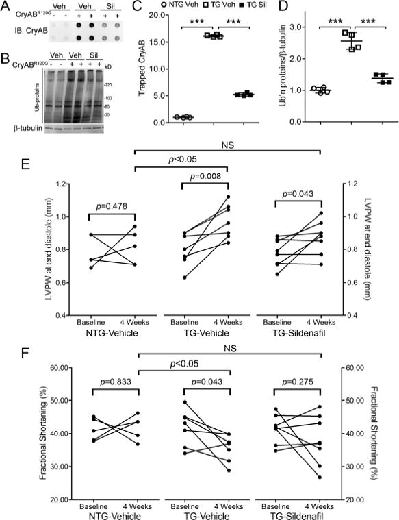
Methods and results: Protein kinase G (PKG) was manipulated genetically and pharmacologically in cultured cardiomyocytes. Activation of PKG increased proteasome peptidase activities, facilitated proteasome-mediated degradation of surrogate (enhanced green fluorescence protein modified by carboxyl fusion of degron CL1) and bona fide (CryAB(R120G)) misfolded proteins, and attenuated CryAB(R120G) overexpression-induced accumulation of ubiquitinated proteins and cellular injury. PKG inhibition elicited the opposite responses. Differences in the abundance of the key 26S proteasome subunits Rpt6 and β5 between the PKG-manipulated and control groups were not statistically significant, but the isoelectric points were shifted by PKG activation. In transgenic mice expressing a surrogate substrate (GFPdgn), PKG activation by sildenafil increased myocardial proteasome activities and significantly decreased myocardial GFPdgn protein levels. Sildenafil treatment significantly increased myocardial PKG activity and significantly reduced myocardial accumulation of CryAB(R120G), ubiquitin conjugates, and aberrant protein aggregates in mice with CryAB(R120G)-based desmin-related cardiomyopathy. No discernible effect on bona fide native substrates of the ubiquitin-proteasome system was observed from PKG manipulation in vitro or in vivo.

Conclusions: PKG positively regulates proteasome activities and proteasome-mediated degradation of misfolded proteins, likely through posttranslational modifications to proteasome subunits. This may be a new mechanism underlying the benefit of PKG stimulation in treating cardiac diseases. Stimulation of PKG by measures such as sildenafil administration is potentially a new therapeutic strategy to treat cardiac proteinopathies.

Keywords: cardiomyopathies; cyclic GMP-dependent protein kinase; desmin; proteasome inhibitors; proteins.


Conflict of interest statement

Conflict of Interest Disclosures: None.

Figures

Figure 1
Figure 1
PKG activation enhances GFPu degradation in cardiomyocytes. Cultured NRVMs were infected with Ad-GFPu and Ad-RFP. Western blot analyses for the steady state protein levels of GFPu and RFP (A, C) were performed using total cell lysates from NRVMs collected at 72h after Ad-PKGcat or Ad-β-gal infection (10MOI, A) or after 48h of sildenafil treatment (1μM, C). In the cycloheximide (CHX) chase assays for GFPu (B, D), CHX treatment (100μM) was started 24h after the infection of Ad-PKGcat/Ad-β-gal (B) or the treatment of sildenafil/DMSO (D). In each run of the CHX chase, the GFPu image density immediately before CHX treatment (i.e., 0 min) was set as an arbitrary unit of one and the GFPu levels of subsequent CHX treated time points were calculated relative to it. A representative western blot image of the CHX chase is shown in the upper section of panels B and D; and GFPu protein decay and half-lives (t1/2) in NRVMs under a given treatment derived from six runs of chase were summarized in the lower section of the panels. *p<0.05 vs. the control group.
Figure 2
Figure 2
Sildenafil stimulates cardiac proteasome proteolytic function in mice. Male GFPdgn mice were subject to two peritoneal injections of sildenafil (10mg/kg) or vehicle control, with a 12-hour interval. Ventricular myocardial samples were collected at 12h after the second injection for the indicated assays. A, Western blot analyses for PKG, total VASP (T-VASP), and Ser-239 phosphorylated VASP (P-VASP). B, Western blot analysis for myocardial GFPdgn protein levels. C, RT-PCR analysis of myocardial GFPdgn mRNA levels. D, Representative fluorescence confocal micrographs of sildenafil or vehicle treated GFPdgn mouse ventricular myocardium. n=6 mice/group.
Figure 3
Figure 3
PKG activation increases proteasome peptidase activities and decreases the pI of proteasome subunits. A, Changes in myocardial proteasome peptidase activities in GFPdgn mice treated with sildenafil as described in Figure 3. Crude protein exacts from ventricular myocardium were used for the indicated peptidase activity assays in presence or absence of ATP. B and C, Effects of PKG activation on proteasome peptidase activities in cultured NRVMs. PKG activation by either forced expression of PKGcat (B) or sildenafil treatment (C) in NRVMs was as described in Figure 1. Crude protein extracts from the cultured cells were used for proteasomal peptidase activity assays in presence or absence of ATP. n=6 biological repeats. NS=not significant, *p<0.05, **p<0.01, ***p<0.005; the same for all subsequent figures. D, Representative images of 2D western blot analyses of Rpt6 and β5 proteasome subunits 72h after Ad-PKGcat or Ad-β-gal infection. Isoelectric focusing gels (IFG) with a pH range from 7 to10 were used for the first dimension separation of proteins based on their pI. For the second dimension separation based on molecular weights, the fully executed IFG was placed in a large center well that was flanked by regular small wells (arrow heads) which were used for one dimensional fractionation of the same source of protein samples as that used for the IFG. The 2D gel was transferred to PVDF membrane and subject to immunoblotting for the indicated proteins.
Figure 4
Figure 4
Activation of PKG enhances proteasome-mediated removal of CryABR120G. NRVMs were cultured for 24h before PKG was manipulated via Ad-PKGcat infection (A, C, F) or sildenafil treatment (B, D, G). Following an additional 24h in culture, NRVMs were infected with either control Ad-β-gal or Ad-HA-CryABR120G. A and B, Representative images of western blot analyses of HA-CryABR120G (upper panel) and β-tubulin in NRVMs 72h after infection with Ad-HA-CryABR120G or Ad-β-gal. C–E, Reduction of steady state HA-CryABR120G protein levels by PKGcat or sildenafil is proteasome-dependent. The proteasome inhibitor bortezomib (BZM, 10nM) or volume corrected saline was applied upon changing of the infection media to normal growth media. NRVMs were harvested after an additional 72 hours for protein extraction. Representative images (C, D) of western blot analyses of the indicated proteins and the pooled densitometry data (E) from four (PKGcat cohort) or three (sildenafil cohort) biological repeats are shown. F and G, CHX chase of HA-CryABR120G and β-tubulin in NRVMs at the indicated time points during PKG activation with PKGcat (F) or sildenafil (G). CHX was administered 24h after Ad-HA-CryABR120G infection. Representative western blot images (upper panel) and the pooled data of the HA-CryABR120G decay and half-life (lower panel) are shown. *p<0.05, **p<0.01 vs. the control group; n= 4 repeats.
Figure 5
Figure 5
PKG regulates the abundance of ubiquitin conjugates in cardiomyocytes overexpressing CryABR120G. A and B, Western blot analyses for total ubiquitinated (Ub’n) proteins in PKG activated NRVMS. NRVMs were cultured and manipulated as described in Figure 4. NRVMs were harvested 72h post Ad-HA-CryABR120G infection. β-Tubulin was probed as loading control. Representative images and a summary of pooled densitometry data from the PKGcat/β-gal cohort (A) and the sildenafil/DMSO cohort (B) are shown in the upper and lower sections of each panel, respectively. C and D, Western blot analyses for total ubiquitinated proteins in PKG inhibited NRVMS. NRVMs were transfected with siPKG/siLuc (C) or treated with KT5823/DMSO (D) 24h after plating, cultured for additional 24h before Ad-HA-CryABR120G or Ad-β-gal infection was performed. The cells were harvested 72h after adenoviral infection.
Figure 6
Figure 6
Sildenafil increases PKG activity and CryABR120G protein degradation in CryABR120G tg mouse hearts. Line 708 CryABR120G tg (+) and Ntg (-) littermate mice were treated with sildenafil (Sil, 10mg/kg/day) or vehicle control (Veh) for 4 weeks via subcutaneous implantation of mini-osmotic pumps. Ventricular myocardium was sampled at the end of treatment for analyses reported here. A, Changes in myocardial PKG activity. B~D, Western blot analyses for PKG, P-VASP, and T-VASP. Representative images (B) and pooled densitometry data (C, D) are presented. E~H, Western blot analyses for CryAB in the soluble (E, G) and insoluble (F, H) fractions. Representative images (E, F) and pooled densitometry data (G, H) are presented.
Figure 7
Figure 7
Sildenafil reduces cardiac aberrant protein aggregation and cardiac hypertrophy and blunts heart function decline in CryABR120G tg mice. The same mouse cohorts as described in Figure 6 were used for data collection here. A and B, The immunoblot images (A) and summary of densitometry data (C) of the filter trap assay for CryAB. The insoluble fraction of myocardial proteins was filtered through a nitrocellulose membrane (pore diameter=0.22μm) and the proteins trapped on the membrane were detected by immunoblotting (IB) for CryAB. B and D, Representative images (B) and a summary of densitometry data (D) of western blot analyses for total ubiquitinated proteins. Each dot represents an individual mouse. E and F, The effect of sildenafil on changes in the end-diastolic left ventricular posterior wall thickness (LVPW) (E) and the fractional shortening (FS) (F) during the 4 weeks of treatment. LVPW and FS data were from serial echocardiographs recorded at 1 day before (baseline) and the final day of (4 weeks) treatment with sildenafil or vehicle (see also online Supplementary Table 1). Two-tailed paired t-tests are used to compare the 4 week time point with the baseline within the group. Inter-group comparisons use one way ANOVA followed by Tukey’s pair-wise tests.
Figure 8
Figure 8
PKG activation protects against proteotoxic stress in cardiomyocytes. PKG activation in NRVMs overexpressing CryABR120G was achieved by PKGcat overexpression (A, C) or sildenafil treatment (B, D), as described in Figure 5. The cultured cells were collected for western blot analyses for caspase 3 and MTT assay and the culture media were simultaneously collected for LDH assays. The LDH/MTT ratio is used to minimize the impact of potential variation resulting from the potential difference in the total cell number on a dish.

Comment in

  • PKG primes the proteasome.
    Gillette TG, Hill JA. Gillette TG, et al. Circulation. 2013 Jul 23;128(4):325-7. doi: 10.1161/CIRCULATIONAHA.113.003955. Epub 2013 Jun 14. Circulation. 2013. PMID: 23770743 No abstract available.

References

    1. Willis MS, Townley-Tilson WH, Kang EY, Homeister JW, Patterson C. Sent to destroy: The ubiquitin proteasome system regulates cell signaling and protein quality control in cardiovascular development and disease. Circ Res. 2010;106:463–478. - PMC - PubMed
    1. Scruggs SB, Zong NC, Wang D, Stefani E, Ping P. Post-translational modification of cardiac proteasomes: Functional delineation enabled by proteomics. Am J Physiol Heart Circ Physiol. 2012;303:H9–18. - PMC - PubMed
    1. Chen Q, Liu JB, Horak KM, Zheng H, Kumarapeli AR, Li J, Li F, Gerdes AM, Wawrousek EF, Wang X. Intrasarcoplasmic amyloidosis impairs proteolytic function of proteasomes in cardiomyocytes by compromising substrate uptake. Circ Res. 2005;97:1018–1026. - PubMed
    1. Tian Z, Zheng H, Li J, Li Y, Su H, Wang X. Genetically induced moderate inhibition of the proteasome in cardiomyocytes exacerbates myocardial ischemia-reperfusion injury in mice. Circ Res. 2012;111:532–542. - PMC - PubMed
    1. Wang X, Pattison JS, Su H. Posttranslational modification and quality control. Circ Res. 2013;112:367–381. - PMC - PubMed

Publication types

MeSH terms