. 2014 May 22;33(21):2807-13.
doi: 10.1038/onc.2013.239. Epub 2013 Jun 17.

Gene amplification of the histone methyltransferase SETDB1 contributes to human lung tumorigenesis

Affiliations
Free PMC article

Gene amplification of the histone methyltransferase SETDB1 contributes to human lung tumorigenesis

M Rodriguez-Paredes et al. Oncogene. .
Free PMC article

Abstract

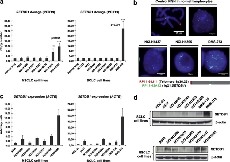
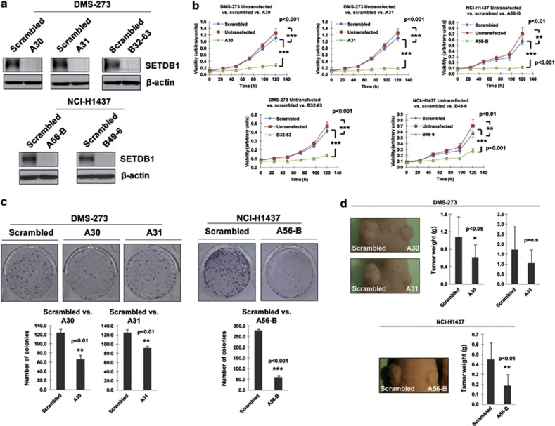
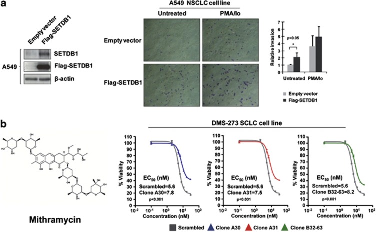
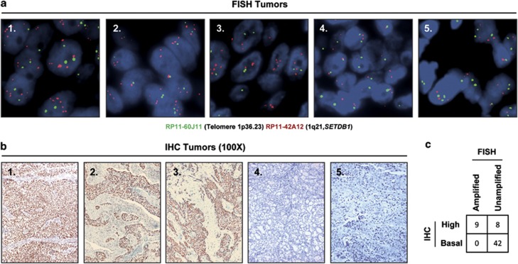
Disruption of the histone modification patterns is one of the most common features of human tumors. However, few genetic alterations in the histone modifier genes have been described in tumorigenesis. Herein we show that the histone methyltransferase SETDB1 undergoes gene amplification in non-small and small lung cancer cell lines and primary tumors. The existence of additional copies of the SETDB1 gene in these transformed cells is associated with higher levels of the corresponding mRNA and protein. From a functional standpoint, the depletion of SETDB1 expression in amplified cells reduces cancer growth in cell culture and nude mice models, whereas its overexpression increases the tumor invasiveness. The increased gene dosage of SETDB1 is also associated with enhanced sensitivity to the growth inhibitory effect mediated by the SETDB1-interfering drug mithramycin. Overall, the findings identify SETDB1 as a bona fide oncogene undergoing gene amplification-associated activation in lung cancer and suggest its potential for new therapeutic strategies.


Figures

Figure 1
Figure 1
Determination of SETDB1 gene amplification and its association with RNA and protein overexpression in lung cancer cell lines. (a) Assessment of SETDB1 copy-number by quantitative genomic PCR. Amplification frequency of SETDB1 (evaluated with SYBR Green, Bio-Rad, Hercules, CA, USA) was calculated by the standard curve method using the 7900HT SDS program. To define an internal control gene, we chose chromosome 1p36.23 because it is the least aneuploid region among our cell lines (PEX19 gene). Primers are available upon request. DNA from normal lung was used as the reference standard. Results are reported as n-fold copy-number increase relative to the PEX19 gene. (b) Fluorescence in situ hybridization for the SETDB1 gene. The UCSC genome browser (http://www.genome.ucsc.edu) was used to select the bacterial artificial chromosome (BAC) clone spanning the 1q21 region for the SETDB1 gene: RP11-42A12. A telomeric BAC clone located in the telomeric 1p36.23 region was used as a control. The BACs were obtained from the BACPAC Resource Center at the Children's Hospital Oakland Research Institute (Oakland, CA, USA). SETDB1 and telomeric probes were labeled with Spectrum Green and Red dUTP (Abbott, Wiesbaden, Germany), respectively, using a CGH Nick Translation Reagent Kit (Abbott Molecular Inc., Des Plaines, IL, USA). The samples were counterstained with 4',6-diamidino-2-phenylindole in Vectashield antifade solution (Burlingame, CA, USA). Gene amplification was observed in the interphases of NCI-H1437, NCI-H1395 and DMS-273. Probes were verified to give a single signal on normal commercial lymphocyte metaphase slides (CGH Reagents, Abbott). Quantitative reverse transcription–PCR (c) and western blot (d) demonstrate higher levels of SETDB1 mRNA and protein (ab12317, Abcam, Cambridge, UK), respectively, in amplified cancer cell lines (H1437, NCI-H1395 and DMS-273) than that in unamplified cells. PCR primers are available upon request.
Figure 2
Figure 2
Growth-promoting effects of SETDB1 in lung cancer. (a) Stable downregulation of the SETDB1 gene by short hairpins using two different target sequences for DMS-273 (clones A30/A31 and clone B32-63) and NCI-H1437 (clones A56-B and B46-9). SETDB1 shRNA sequences are available upon request. (b) The short hairpin SETDB1-depleted cells were less viable in the 3-(4,5-dimethyl-2-thiazolyl)-2,5-diphenyl-2H-tetrazolium bromide (MTT) assay than in the untransfected or scrambled shRNA-transfected cells (P-values obtained by the analysis of variance (ANOVA) test). (c) The colony formation assay showed that DMS-273 and NCI-H1437 cells stably transfected with the shRNA against SETDB1 formed significantly fewer colonies than scrambled shRNA-transfected cells (P-values obtained by the ANOVA test). Data shown are means±s.d., n=3. (d) Effect of SETDB1 shRNA-mediated depletion on the growth of DMS-273 and NCI-H1437 xenografts in nude mice. Tumor volume was monitored over time and the tumor was excised and weighed at 30 days. There was a significant decrease in tumor weight in the SETDB1 shRNA-stably transfected cells (P-values obtained by the ANOVA test). Data shown are means±s.d., n=10.
Figure 3
Figure 3
Impact of SETDB1 on invasiveness and chemosensitivity. (a) Effect of SETDB1 on the invasion potential of A549 cells determined by the matrigel invasion assay. Cells were transfected with 3 μg of Flag-SETDB1 or empty vector in 60 mm dishes. After 24 h, cells were stimulated or not with phorbol myristate acetate (PMA) plus ionomycin (Io) for 30 min. Then cells were trypsinized, and 5 × 10⁴ cells were resuspended in serum-free media and added to the upper compartment of a transwell coated with 1 mg/ml Matrigel (BD Biosciences, Lexington, KY, USA). Media with 10% fetal bovine serum was added in the lower compartment and cells were incubated at 37 °C for 42 h. Invasive cells were fixed with phosphate-buffered saline 4% paraformaldehide, stained with 0.5% violet crystal and visualized and photographed under a × 10 magnification objective with a microscope. Invasive cells were counted using ImageJ 1.45s (Wayne Rasband, National Institutes of Health, Bethesda, MD, USA) and percentage of invasive cells were represented. Results are the mean of at least three experiments by duplicate and the significance was determined using analysis of variance test. *<P=0.05. (b) Cancer cells harboring the SETDB1 gene amplification are sensitive to the decrease in cell viability caused by mithramycin, a SETDB1-interfering drug. 3-(4,5-dimethyl-2-thiazolyl)-2,5-diphenyl-2H-tetrazolium bromide (MTT) assays in control-scrambled shRNA DMS-273-transfected cells in comparison with three shRNA-stable downregulated SETDB1 clones (A21, A30 and A31) show enhanced inhibition of viability in cells with SETDB1 gene amplification-mediated overexpression.
Figure 4
Figure 4
Detection of SETDB1 gene amplification and its associated overexpression in primary tumors from lung cancer patients. (a) Fluorescence in situ hybridization for the SETDB1 gene shows gene amplification in the primary lung tumors 1, 2 and 3. SETDB1 unamplified tumors are shown in the cases 4 and 5. The UCSC genome browser (http://www.genome.ucsc.edu) was used to select the bacterial artificial chromosome (BAC) clone RP11-42A12 spanning the 1q21 region of SETDB1 gene. A telomeric BAC clone located in the telomeric 1p36.23 region was used as a control. (b) Immunohistochemistry for SETDB1 (HPA018142, Sigma-Aldrich, St Louis, MO, USA) shows overexpression of the protein in the above shown three primary lung tumors harboring SETDB1 gene amplification. Minimal expression is detected in the unamplified cases (4 and 5). Magnification × 100. (c) Association between SETDB1 gene amplification and overexpression in the studied fifty-nine cases is shown. Fisher's test, two-tailed P-value<0.0001.

References

    1. Jones PA, Baylin SB. The epigenomics of cancer. Cell. 2007;128:683–692. - PMC - PubMed
    1. Berdasco M, Esteller M. Aberrant epigenetic landscape in cancer: how cellular identity goes awry. Dev Cell. 2010;19:698–711. - PubMed
    1. Füllgrabe J, Kavanagh E, Joseph B. Histone onco-modifications. Oncogene. 2011;30:3391–3403. - PubMed
    1. Fraga MF, Ballestar E, Villar-Garea A, Boix-Chornet M, Espada J, Schotta G, et al. Loss of acetylation at Lys16 and trimethylation at Lys20 of histone H4 is a common hallmark of human cancer. Nat Genet. 2005;37:391–400. - PubMed
    1. Seligson DB, Horvath S, Shi T, Yu H, Tze S, Grunstein M, et al. Global histone modification patterns predict risk of prostate cancer recurrence. Nature. 2005;435:1262–1266. - PubMed

Publication types

MeSH terms

Substances