. 2013:4:1983.
doi: 10.1038/ncomms2983.

A molecular explanation for the recessive nature of parkin-linked Parkinson's disease

Affiliations
Free PMC article

A molecular explanation for the recessive nature of parkin-linked Parkinson's disease

Donald E Spratt et al. Nat Commun. 2013.
Free PMC article

Abstract

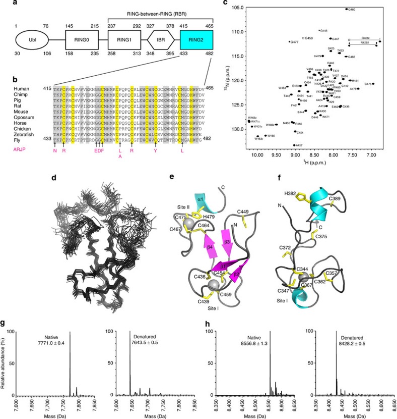
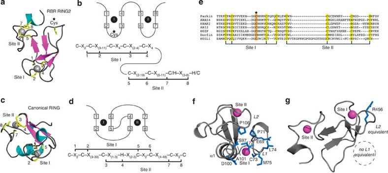
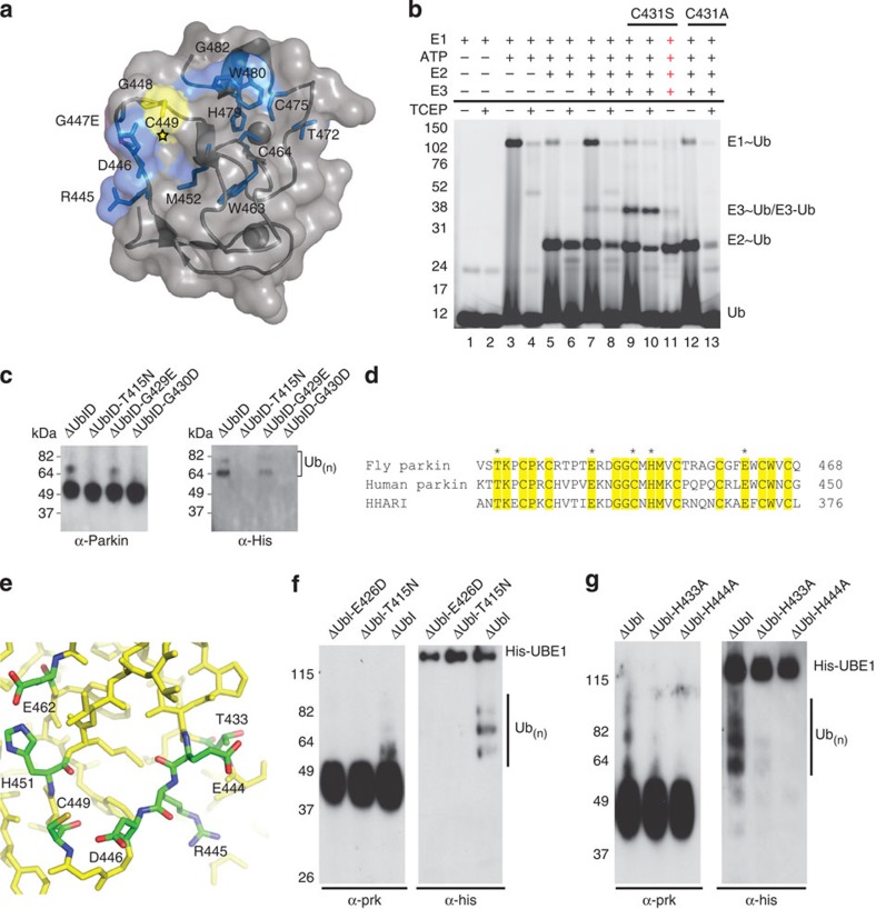
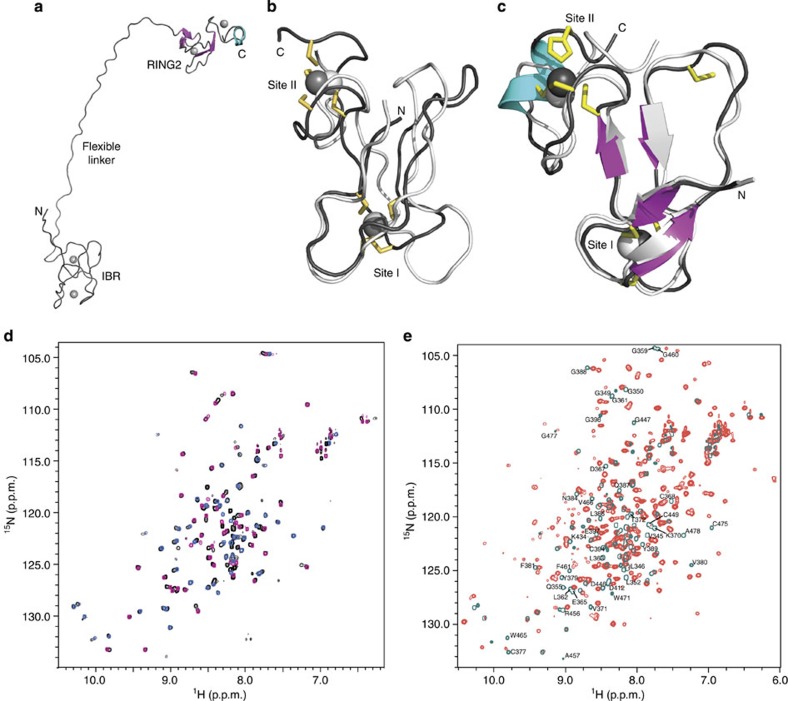
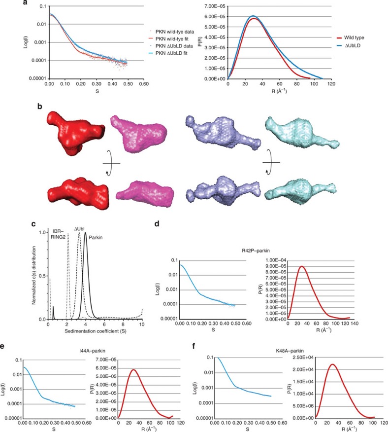
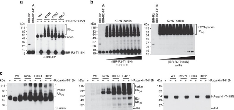
Mutations in the park2 gene, encoding the RING-inBetweenRING-RING E3 ubiquitin ligase parkin, cause 50% of autosomal recessive juvenile Parkinsonism cases. More than 70 known pathogenic mutations occur throughout parkin, many of which cluster in the inhibitory amino-terminal ubiquitin-like domain, and the carboxy-terminal RING2 domain that is indispensable for ubiquitin transfer. A structural rationale showing how autosomal recessive juvenile Parkinsonism mutations alter parkin function is still lacking. Here we show that the structure of parkin RING2 is distinct from canonical RING E3 ligases and lacks key elements required for E2-conjugating enzyme recruitment. Several pathogenic mutations in RING2 alter the environment of a single surface-exposed catalytic cysteine to inhibit ubiquitination. Native parkin adopts a globular inhibited conformation in solution facilitated by the association of the ubiquitin-like domain with the RING-inBetweenRING-RING C-terminus. Autosomal recessive juvenile Parkinsonism mutations disrupt this conformation. Finally, parkin autoubiquitinates only in cis, providing a molecular explanation for the recessive nature of autosomal recessive juvenile Parkinsonism.


Figures

Figure 1
Figure 1. Sequence alignment, structure and metal analysis of parkin RING2 domain.
(a) Domain structure of parkin showing the C-terminal RING1, IBR and RING2 domains found in all RBR E3 ligase proteins. The ubiquitin-like (Ubl) and RING0 domains are specific to the parkin E3 ligase. Residue numbering is shown for both the human (top) and D. melanogaster (bottom) parkin sequences. (b) Multiple sequence alignment of the RING2 domain for parkin orthologues. Sequence numbers are indicated for the human and D. melanogaster species only. Conserved (grey) and cysteine/histidine (yellow) residues are highlighted. Substitutions that contribute to ARJP in the parkin RING2 domain are shown below the sequences (magenta). (c) Assigned 600 MHz 1H–15N HSQC spectrum of 13C,15N-labelled parkin RING2 (500 μM in 20 mM Tris–HCl, 120 mM NaCl, 1 mM DTT, pH 7.25), labelled using the one-letter amino acid code and residue number according to the D. melanogaster parkin sequence. (d) Superposition of the 20 lowest energy solution structures of parkin RING2 (residues 430–482, backbone r.m.s.d. 0.82±0.17 Å). (e) Ribbon structure of parkin RING2 showing β-strands β1 (T433–P435), β2 (P442–E444), β3 (H451–V453) and β4 (E462–C464), and helix α1 (M476–W480). Side chains for Zn2+-coordinating residues are shown in yellow. (f) Structure of the HHARI RING2 (ref. 35) (PDB accession code 1WD2) showing different zinc occupancy and fold compared with parkin (e). Deconvoluted mass spectra for native and denatured parkin RING2 (g), and native and denatured HHARI RING2 (h). The mass differences of 127.5 Da (g) and 128.4 Da (h) indicate the presence of two bound Zn2+ ions in the native proteins. Raw data are found in Supplementary Figure S2. Structures were visualized using Pymol (PyMOL Molecular Graphics System, Version 1.5.0.4 Schrödinger, LLC).
Figure 2
Figure 2. RING2 domain of parkin and other RBR E3 ligases do not recruit E2 enzymes.
Parkin RING2 defines a novel structure common to RBR ubiquitin ligases that are unable to recruit E2 enzymes. The structures and schematic representations of (a,b) RBR RING2 (parkin) and (c,d) RING (TRAF6, PDB 3HCS) domains showing differences in Zn2+ coordination and consensus sequences. (e) Alignment of the RING2 domains of RBR ligases showing residues predicted to coordinate Zn2+ (yellow) based on the parkin RING2 structure and the proposed catalytic cysteine conserved in all sequences (orange). (f) Key residues and elements used for E2 recruitment by the canonical RING E3 ligase TRAF6 are found in two loops (L1, L2) formed through Zn2+ coordination that are absent in (g) parkin RING2. The structures are oriented using the loops involved in Zn2+ coordination for TRAF6 site II and parkin RING2 site I, which exhibit the closest structural similarity.
Figure 3
Figure 3. Pathogenic mutations in parkin RING2.
The structure of parkin predicts catalytic residues conserved between parkin and HHARI. (a) The structure of parkin with the catalytic cysteine highlighted (star) and chemical-shift perturbation experiments show affected residues (blue) in parkin RING2 by the ARJP substitution G447E (magenta). (b) The formation of a reducible parkin~Ub thiolester was monitored using His-SUMO-IBR-R2-parkin. Lanes 1–2 contain E1, Mg2+ and Cy5–ubiquitin incubated for 10 min at 37 oC. Lanes 3–4 have ATP added for 10 min. Lanes 5–6 have E2 added followed by a further 10-min incubation. Lanes 7–8 have wild-type parkin added. Lanes 9–11 have C431S–parkin added, with NaOH added to lane 11, indicated by red crosses. Lanes 12–13 have C431A–parkin added; ‘−’ and ‘+’ below the line indicate the absence or presence of TCEP. (c) An autoubiquitination assay of folded pathogenic mutants. Parkin–ubiquitin conjugates are detected by western blotting using parkin (left) and His-ubiquitin (right) antibodies. (d) An alignment of the RING2 domains of fly and human parkin, and HHARI. Conserved residues are shaded yellow and the potential catalytic residues are marked with an asterisk. (e) Close-up view of the surface of parkin RING2 with the potential catalytic residues indicated. (f) Autoubiquitination assay of active parkin (ΔUbl) and parkin ΔUbl-E426D. (g) Autoubiquitination assays of active parkin (ΔUbl) and parkin ΔUbl-H433A and ΔUbl-H444A. In f and g, parkin–ubiquitin conjugates are detected by western blotting using parkin (left) and His-ubiquitin (right) antibodies.
Figure 4
Figure 4. The structure of fly IBR–RING2 shows the domains are remote in parkin but interact with RING0 and RING1.
(a) Representative ribbon structure of parkin IBR–RING2 showing the flexible linker between the IBR and RING2 domains. (b) The superposition of parkin IBR domains from human (white, PBD accession code 2JMO) and fly (black). (c) The superposition of RING2 domains from fly parkin RING2417–482 (white) and fly parkin IBR–RING2342–482 (coloured). The two domains adopt similar folds, although the IBR domains do not contain formal β-strands. (d) Superposition of 1H–15N HSQC spectra for the fly IBR–RING2 (black contours) with spectra for the individual fly parkin IBR342–402 (pink) and RING2417–482 (blue) domains. (e) Superposition of 1H–15N HSQC spectra for parkin RING0–RING1–IBR–RING2 (red contours) and IBR–RING2342–482 (teal contours). The large number of chemical-shift changes indicates that RING0 and RING1 interact with IBR–RING2. Residues in both IBR and RING2 that undergo the largest changes in chemical shift are indicated near their positions from the IBR–RING2 assignment.
Figure 5
Figure 5. The tertiary structure of parkin is maintained by the Ubl.
(a) Superposition of the scattering data (left) and distance distribution (right) plots from purified human parkin samples, wild-type (red) and ΔUblD–parkin (blue). The plots show the quality of the data and the radius of gyration. (b) Representative and averaged ab initio models of wild-type parkin (red/pink) and ΔUbl–parkin (blue/cyan). Two views for each protein are shown rotated 90o about the x axis. (c) Sedimentation velocity experiments of parkin, ΔUblD–parkin, and IBR–RING2. All data were analysed using the Lamm equation and fit to a c(s) distribution. Sedimentation coefficients, corrected to 20 °C and in H2O, were determined to be 4.1 S for full-length parkin, 3.5 S for ΔUblD–parkin and 2.2 S for IBR–RING2. Fitted frictional ratios (f/f0) were calculated to be 1.38 for full-length parkin, 1.53 for ΔUbl–parkin and 1.31 for IBR–RING2. Sedimentation velocity experiments were performed at 20 °C using 10–16 μM protein in 25 mM Tris-HCl, 50 mM NaCl, 0.5 mM TCEP, pH 8.0. (d) Scattering data (left) and distance distribution (right) plots for R42P–parkin, (e) I44A–parkin, and (f) K48A–parkin.
Figure 6
Figure 6. Parkin is not a substrate of parkin in trans.
(a) Ubiquitination assay using IBR–RING2–T415N (IBR–R2–T415N) as a substrate. Each parkin species point mutation is assayed in the absence (−) or presence (+) of substrate. The first lane shows wild-type active IBR–RING2, the second lane shows the inactive mutant. Parkin–ubiquitin conjugates are detected by western blotting using a parkin antibody. (b) K27N–parkin is assayed for activity towards IBR-R2-T415N with increasing substrate concentration. Parkin–ubiquitin conjugates are detected by western blotting using parkin (left) and His-ubiquitin (right) antibodies. (c) Ubiquitination assay using full-length HA–parkin–T415N as a substrate. Parkin–ubiquitin conjugates are detected by western blotting using parkin and His-ubiquitin antibodies. Unmodified HA-parkin-T415N is detected using HA antibodies. The asterisk denotes a non-specific band.

References

    1. Kitada T. et al. Mutations in the parkin gene cause autosomal recessive juvenile parkinsonism. Nature 392, 605–608 (1998). - PubMed
    1. Lucas J. I., Arnau V. & Marin I. Comparative genomics and protein domain graph analyses link ubiquitination and RNA metabolism. J. Mol. Biol. 357, 9–17 (2006). - PubMed
    1. Marin I., Lucas J. I., Gradilla A. C. & Ferrus A. Parkin and relatives: the RBR family of ubiquitin ligases. Physiol. Genomics 17, 253–263 (2004). - PubMed
    1. Narendra D., Kane L. A., Hauser D. N., Fearnley I. M. & Youle R. J. p62/SQSTM1 is required for Parkin-induced mitochondrial clustering but not mitophagy; VDAC1 is dispensable for both. Autophagy 6, 1090–1106 (2010). - PMC - PubMed
    1. Ziviani E., Tao R. N. & Whitworth A. J. Drosophila parkin requires PINK1 for mitochondrial translocation and ubiquitinates mitofusin. Proc. Natl Acad. Sci. USA 107, 5018–5023 (2010). - PMC - PubMed

Publication types

Substances