Comparative Study
. 2014 Jan;134(1):87-95.
doi: 10.1038/jid.2013.269. Epub 2013 Jun 14.

Dominant Th1 and minimal Th17 skewing in discoid lupus revealed by transcriptomic comparison with psoriasis

Affiliations
Comparative Study

Dominant Th1 and minimal Th17 skewing in discoid lupus revealed by transcriptomic comparison with psoriasis

Ali Jabbari et al. J Invest Dermatol. 2014 Jan.

Erratum in

  • J Invest Dermatol. 2014 Jun;134(6):1780

Abstract

Discoid lupus erythematosus (DLE) is the most common skin manifestation of lupus. Despite its high frequency in systemic lupus in addition to cases without extracutaneous manifestations, targeted treatments for DLE are lacking, likely because of a dearth of knowledge of the molecular landscape of DLE skin. Here, we profiled the transcriptome of DLE skin in order to identify signaling pathways and cellular signatures that may be targeted for treatment purposes. Further comparison of the DLE transcriptome with that of psoriasis, a useful reference given our extensive knowledge of molecular pathways in this disease, provided a framework to identify potential therapeutic targets. Although a growing body of data support a role for IL-17 and T helper type 17 (Th17) cells in systemic lupus, we show a relative enrichment of IFN-γ-associated genes without that for IL-17-associated genes in DLE. Extraction of T cells from the skin of DLE patients identified a predominance of IFN-γ-producing Th1 cells and an absence of IL-17-producing Th17 cells, complementing the results from whole-skin transcriptomic analyses. These data therefore support investigations into treatments for DLE that target Th1 cells or the IFN-γ signaling pathway.


Figures
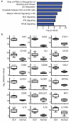
Figure 1
Figure 1
Transcriptomic analysis of DLE. (A) Selected top canonical pathways from interrogation of the DLE (n=7) versus normal transcriptome (n=13) with Ingenuity Pathway Analysis. (B) Quantitative RT-PCR of selected upregulated genes in the DLE versus normal transcriptome corresponding to interferon-associated genes (MX1, OASL, STAT1), immune cell associated genes, and selected cytokines in DLE (n=7) and normal (n=6) samples. Genes were normalized to human ribosomal acidic protein. Box and whisker plots (middle line: median; box: lower to upper quartile; whiskers: minimum and maximum), * p < 0.05, ** p < 0.005, *** p < 5 × 10⁻⁴, **** p < 5 × 10⁻⁵, † p < 5 × 10⁻⁶, †† p < 5 × 10⁻⁷, †††† p < 5 × 10⁻⁹
Figure 2
Figure 2
Microscopic and immunohistologic comparison of DLE and psoriasis. Skin sections from patients with DLE, psoriasis, or healthy control patients were stained with hematoxylin and eosin or were subjected to immunohistochemistry with antibodies specific for CD3, CD11c, or CD8. Scale bar = 100 μm.
Figure 3
Figure 3
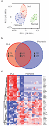
Transcriptomic comparison between DLE and psoriasis. (A) Principal components analysis plot comparing DLE (n=7), psoriasis (PsO, n=18), and normal control (n=13) samples. (B) Venn diagram comparing upregulated and downregulated genes from DLE versus normal skin (DLE) and psoriasis versus normal skin (PsO). (C) Heat map of the most upregulated and downregulated genes in DLE versus psoriasis comparison.
Figure 4
Figure 4
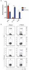
Comparison of T helper subsets in DLE and psoriasis. (A) Quantitative RT-PCR comparison of T helper subset-associated cytokines in DLE and psoriasis (PsO). * p < 0.05, ** p < 5 × 10⁻⁵, † p < 5 × 10⁻⁶. (B) Intracellular cytokine staining of CD4 T cells extracted from lesions from patients with DLE (n=4) or psoriasis (n=4) for IFN-γ or IL-17A, stimulated with (+ PMA/I, right column) or without (− PMA/I, left column) PMA and ionomycin. Numbers are percentages of CD3+CD4+ events positive for either IFN-γ or IL-17A.

References

    1. Alunno A, Bartoloni E, Bistoni O, Nocentini G, Ronchetti S, Caterbi S, et al. Balance between Regulatory T and Th17 Cells in Systemic Lupus Erythematosus: The Old and the New. Clin Dev Immunol. 2012;2012:823085. - PMC - PubMed
    1. Baechler EC, Batliwalla FM, Karypis G, Gaffney PM, Ortmann WA, Espe KJ, et al. Interferon-inducible gene expression signature in peripheral blood cells of patients with severe lupus. Proc Natl Acad Sci U S A. 2003;100:2610–5. - PMC - PubMed
    1. Balanescu P, Balanescu E, Tanasescu C, Nicolau A, Tanasescu R, Grancea C, et al. T helper 17 cell population in lupus erythematosus. Rom J Intern Med. 2010;48:255–9. - PubMed
    1. Bauer JW, Baechler EC, Petri M, Batliwalla FM, Crawford D, Ortmann WA, et al. Elevated serum levels of interferon-regulated chemokines are biomarkers for active human systemic lupus erythematosus. PLoS Med. 2006;3:e491. - PMC - PubMed
    1. Bennett L, Palucka AK, Arce E, Cantrell V, Borvak J, Banchereau J, et al. Interferon and granulopoiesis signatures in systemic lupus erythematosus blood. J Exp Med. 2003;197:711–23. - PMC - PubMed

Publication types

Associated data