

Perfusion and diffusion MRI combined with ¹¹C-methionine PET in the preoperative evaluation of suspected adult low-grade gliomas
- PMID: 23771511
- PMCID: PMC3742413
- DOI: 10.1007/s11060-013-1178-3
Perfusion and diffusion MRI combined with ¹¹C-methionine PET in the preoperative evaluation of suspected adult low-grade gliomas
Abstract
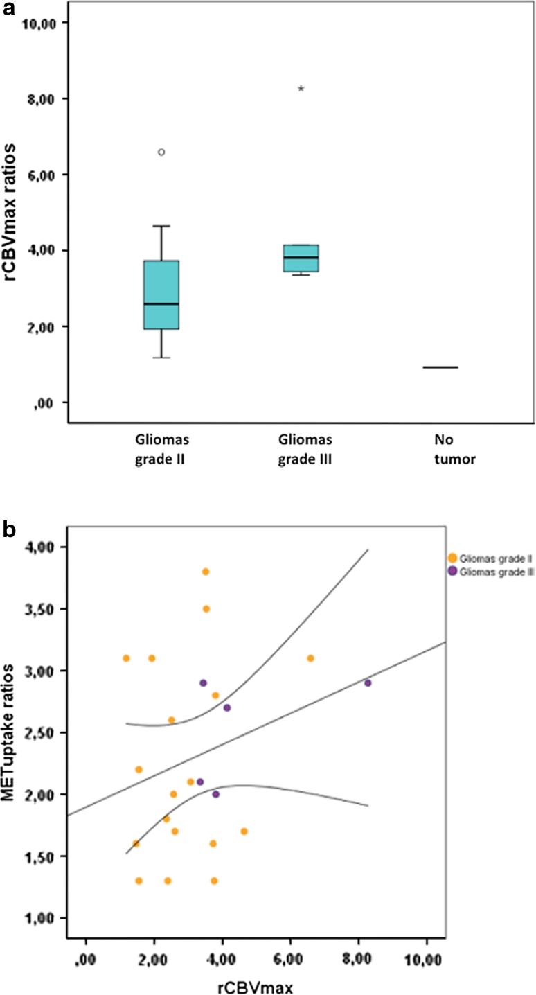
Perfusion and diffusion magnetic resonance imaging (pMRI, dMRI) are valuable diagnostic tools for assessing brain tumors in the clinical setting. The aim of this study was to determine the correlation of pMRI and dMRI with ¹¹C-methionine positron emission tomography (MET PET) in suspected low-grade gliomas (LGG) prior to surgery. Twenty-four adults with suspected LGG were enrolled in an observational study and examined by MET PET, pMRI and dMRI. Histological tumor diagnosis was confirmed in 23/24 patients (18 gliomas grade II, 5 gliomas grade III). The maximum relative cerebral blood volume (rCBV(max)) and the minimum mean diffusivity (MD(min)) were measured in tumor areas with highest MET uptake (hotspot) on PET by using automated co-registration of MRI and PET scans. A clearly defined hotspot on PET was present in all 23 tumors. Regions with rCBV(max) corresponded with hotspot regions in all tumors, regions with MD(min) corresponded with hotspot regions in 20/23 tumors. The correlation between rCBV(max) (r = 0.19, P = 0.38) and MD(min) (r = -0.41, P = 0.053) with MET uptake in the hotspot was not statistically significant. Taken into account the difficulties of measuring perfusion abnormalities in non-enhancing gliomas, this study demonstrates that co-registered MET PET and pMRI facilitates the identification of regions with rCBV(max). Furthermore, the lack of a clear positive correlation between tumor metabolism in terms of MET uptake and tumor vascularity measured as rCBV(max) suggests that combined pMRI/PET provides complementary baseline imaging data in these tumors.
Figures


References
-
- Lote K, Egeland T, Hager B, Stenwig B, Skullerud K, Berg-Johnsen J, Storm-Mathisen I, Hirschberg H. Survival, prognostic factors, and therapeutic efficacy in low-grade glioma: a retrospective study in 379 patients. J Clin Oncol. 1997;15:3129–3140. - PubMed
-
- Soffietti R, Baumert BG, Bello L, von Deimling A, Duffau H, Frenay M, Grisold W, Grant R, Graus F, Hoang-Xuan K, Klein M, Melin B, Rees J, Siegal T, Smits A, Stupp R, Wick W. Guidelines on management of low-grade gliomas: report of an EFNS-EANO* task force. Eur J Neurol. 2010;17:1124–1133. doi: 10.1111/j.1468-1331.2010.03151.x. - DOI - PubMed
Publication types
MeSH terms
Substances
LinkOut - more resources
Full Text Sources
Other Literature Sources
Medical
Miscellaneous
