. 2013 Aug 15;305(4):L291-300.
doi: 10.1152/ajplung.00094.2013. Epub 2013 Jun 14.

Conditional deletion of FAK in mice endothelium disrupts lung vascular barrier function due to destabilization of RhoA and Rac1 activities

Affiliations

Conditional deletion of FAK in mice endothelium disrupts lung vascular barrier function due to destabilization of RhoA and Rac1 activities

Tracy Thennes Schmidt et al. Am J Physiol Lung Cell Mol Physiol. .

Abstract

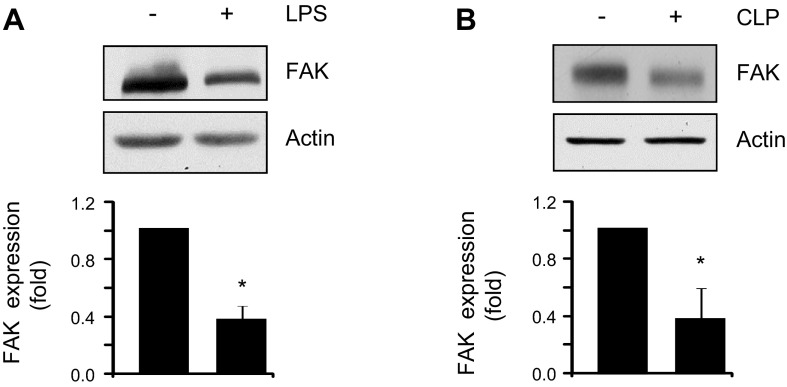
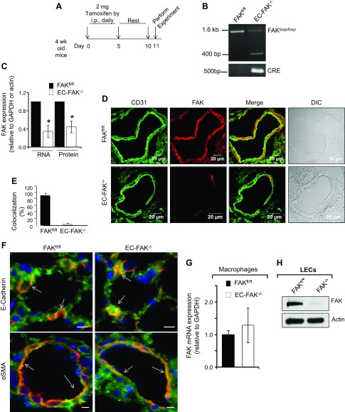
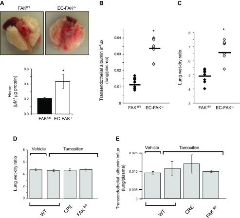
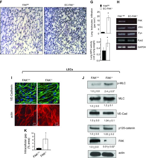
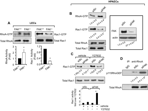
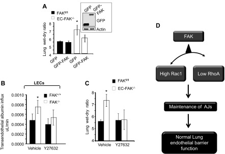
Loss of lung-fluid homeostasis is the hallmark of acute lung injury (ALI). Association of catenins and actin cytoskeleton with vascular endothelial (VE)-cadherin is generally considered the main mechanism for stabilizing adherens junctions (AJs), thereby preventing disruption of lung vascular barrier function. The present study identifies endothelial focal adhesion kinase (FAK), a nonreceptor tyrosine kinase that canonically regulates focal adhesion turnover, as a novel AJ-stabilizing mechanism. In wild-type mice, induction of ALI by intraperitoneal administration of lipopolysaccharide or cecal ligation and puncture markedly decreased FAK expression in lungs. Using a mouse model in which FAK was conditionally deleted only in endothelial cells (ECs), we show that loss of EC-FAK mimicked key features of ALI (diffuse lung hemorrhage, increased transvascular albumin influx, edema, and neutrophil accumulation in the lung). EC-FAK deletion disrupted AJs due to impairment of the fine balance between the activities of RhoA and Rac1 GTPases. Deletion of EC-FAK facilitated RhoA's interaction with p115-RhoA guanine exchange factor, leading to activation of RhoA. Activated RhoA antagonized Rac1 activity, destabilizing AJs. Inhibition of Rho kinase, a downstream effector of RhoA, reinstated normal endothelial barrier function in FAK-/- ECs and lung vascular integrity in EC-FAK-/- mice. Our findings demonstrate that EC-FAK plays an essential role in maintaining AJs and thereby lung vascular barrier function by establishing the normal balance between RhoA and Rac1 activities.

Keywords: acute lung injury; adherens junctions; endothelial barrier; focal adhesion kinase.


Figures

Fig. 1.
Fig. 1.
Focal adhesion kinase (FAK) protein expression is decreased during acute lung injury in mice. Lung lysates from wild-type mice exposed to vehicle or lipopolysaccharide (LPS, 30 mg/kg, 6 h) (A) or subjected to cecal ligation (control) or cecal ligation and puncture (CLP, 22 h) (B) were immunoblotted with anti-FAK or anti-β-actin (loading control) antibodies (Abs). Bar graphs show the densitometric analysis of FAK expression from 4 lungs/group (A and B); *statistically significant difference from vehicle or control lungs (P < 0.05).
Fig. 2.
Fig. 2.
Inducible deletion of endothelial FAK. A: schematic of tamoxifen-induced deletion of FAK in CRE-expressing FAKfl/fl mice. Four-week-old FAKfl/fl (control)- and FAKfl/fl-expressing Cre mice were injected with 2 mg tamoxifen ip for 5 consecutive days, followed by a rest period of 5 days. Mice were used for experiments on the 11th day. B: genotype showing tamoxifen deletion of FAK. Tails from FAKfl/fl or endothelial cell (EC)-FAK−/− mice were digested, and FAK and Cre expression was determined using specific primers. Cre induces FAK deletion in Cre-expressing EC-FAK−/− mice that is absent in FAKfl/fl mice. C–E: assessment of endothelial FAK expression. Lungs harvested from FAKfl/fl or EC-FAK−/− mice were homogenized, and FAK mRNA and protein expression was quantified using FAK primers and FAK antibody (C). The bar graph shows mean ± SD of FAK expression normalized against the housekeeping gene GAPDH (for RNA) or β-actin (for protein). *Statistically significant difference from FAKfl/fl lungs (P < 0.05, n = 4–5). D: FAKfl/fl and EC-FAK−/− lung sections were immunostained with anti-FAK (red) and anti-CD31 (green) Abs, followed by appropriate Alexa-fluor-conjugated secondary Abs to assess FAK expression in CD31+ endothelial cells. Scale bar, 20 μM. Bar graph shows %CD31+ endothelial cells that express FAK in EC-FAK null and FAKfl/fl lungs from 6–7 individual vessels from multiple lung sections (E). F: lung sections from FAKfl/fl and EC-FAK−/− mice were either coimmunostained with anti-FAK (green) and anti-E-cadherin (red) or anti-FAK and anti-α-smooth muscle actin (α-SMA; red) Abs to confirm FAK expression in epithelial and smooth muscle cells, respectively. Arrows show similar FAK colocalization with E-cadherin or α-SMA in FAKfl/fl and EC-FAK−/− mice lungs. Scale bars, 10 μM. Images represent 3–4 mice/group. G: macrophages were isolated from bronchoalveloar lavage (BAL) of FAKfl/fl and EC-FAK−/− mice after which RNA was extracted. FAK expression was quantified using suitable primers as described in materials and methods. GAPDH was used as an internal control. Plot represents the mean ± SD from 4 mice. H: lung endothelial cells (LECs) isolated from FAKfl/fl mice infected with β-gal (control, FAK+/+) or Cre adenovirus (FAK−/−) were lysed to assess FAK expression using actin as a loading control. Blot is representative of five individual isolations.
Fig. 3.
Fig. 3.
Endothelial FAK deletion disrupts lung vascular barrier function. A: loss of EC-FAK leads to diffuse lung hemorrhage. Top, representative images of buffer-perfused lungs harvested from EC-FAK−/− and FAKfl/fl mice. Bottom, mean ± SD of heme concentration from four FAKfl/fl and EC-FAK−/− mouse lungs. *Values significantly different from FAKfl/fl lungs (P < 0.05). B and C: EC-FAK deletion increases lung microvascular permeability. Transendothelial albumin influx into lung parenchyma (B) and lung wet-to-dry weight ratio (C) were determined as described in materials and methods to quantify lung vascular protein permeability and lung edema formation, respectively. *Significant difference from FAKfl/fl lungs (P < 0.05; n = 7–8 mice/group). D and E: tamoxifen does not affect lung vascular permeability or lung edema formation in wild-type (WT), CRE, or FAKfl/fl mice. Tamoxifen was injected into WT, CRE, or FAKfl/fl mice following the protocol described in Fig. 1A. Mice were killed at day 11 to determine lung wet-to-dry weight ratios (D) or transendothelial albumin influx into lung parenchyma (E). Data represent means ± SD from three groups of four lungs. F and G: EC-FAK deletion induces lung leukocyte infiltration and activation. Representative images of hematoxylin and eosin (H & E)-stained lung sections from multiple FAKfl/fl and EC-FAK−/− mice (F). Scale bars, 20 μm. G: top, plot shows mean ± SD leukocyte infiltration in the lungs. *Significant difference from FAKfl/fl lungs (P < 0.05, n = 4 mice/group). Bottom, lungs from FAKfl/fl or EC-FAK−/− were harvested, and myeloperoxidase (MPO) activity was quantified as indicated in materials and methods. *Significant difference from FAKfl/fl lungs (P < 0.05, n = 4 mice/group). H: endothelial FAK deletion does not alter mRNA expression of related kinases. RNA was extracted from FAKfl/fl and EC-FAK−/− mouse lungs. The expression of cSrc, Fyn, Pyk2, and FAK was analyzed by RT-PCR using specific primers. GAPDH was used as a loading control. Results represent data from three individual lungs. I–K: FAK deletion disrupts adherens junctions. FAK+/+ and FAK−/− LECs were immunostained with anti-vascular endothelial (VE)-cadherin (green) antibody or rhodamine phallodin (red) as described in materials and methods (I). Immunoblot from FAK+/+ and FAK−/− lysates was probed with indicated Abs (J). *Significant difference between FAK+/+ vs. FAK−/− LECs (n = 3; P < 0.05). K: plot shows mean ± SD of interendothelial gap area in FAK+/+ and FAK−/− monolayers. *Significant difference from FAK+/+ monolayers (P < 0.05, n = 5).
Fig. 3.
Fig. 3.
Endothelial FAK deletion disrupts lung vascular barrier function. A: loss of EC-FAK leads to diffuse lung hemorrhage. Top, representative images of buffer-perfused lungs harvested from EC-FAK−/− and FAKfl/fl mice. Bottom, mean ± SD of heme concentration from four FAKfl/fl and EC-FAK−/− mouse lungs. *Values significantly different from FAKfl/fl lungs (P < 0.05). B and C: EC-FAK deletion increases lung microvascular permeability. Transendothelial albumin influx into lung parenchyma (B) and lung wet-to-dry weight ratio (C) were determined as described in materials and methods to quantify lung vascular protein permeability and lung edema formation, respectively. *Significant difference from FAKfl/fl lungs (P < 0.05; n = 7–8 mice/group). D and E: tamoxifen does not affect lung vascular permeability or lung edema formation in wild-type (WT), CRE, or FAKfl/fl mice. Tamoxifen was injected into WT, CRE, or FAKfl/fl mice following the protocol described in Fig. 1A. Mice were killed at day 11 to determine lung wet-to-dry weight ratios (D) or transendothelial albumin influx into lung parenchyma (E). Data represent means ± SD from three groups of four lungs. F and G: EC-FAK deletion induces lung leukocyte infiltration and activation. Representative images of hematoxylin and eosin (H & E)-stained lung sections from multiple FAKfl/fl and EC-FAK−/− mice (F). Scale bars, 20 μm. G: top, plot shows mean ± SD leukocyte infiltration in the lungs. *Significant difference from FAKfl/fl lungs (P < 0.05, n = 4 mice/group). Bottom, lungs from FAKfl/fl or EC-FAK−/− were harvested, and myeloperoxidase (MPO) activity was quantified as indicated in materials and methods. *Significant difference from FAKfl/fl lungs (P < 0.05, n = 4 mice/group). H: endothelial FAK deletion does not alter mRNA expression of related kinases. RNA was extracted from FAKfl/fl and EC-FAK−/− mouse lungs. The expression of cSrc, Fyn, Pyk2, and FAK was analyzed by RT-PCR using specific primers. GAPDH was used as a loading control. Results represent data from three individual lungs. I–K: FAK deletion disrupts adherens junctions. FAK+/+ and FAK−/− LECs were immunostained with anti-vascular endothelial (VE)-cadherin (green) antibody or rhodamine phallodin (red) as described in materials and methods (I). Immunoblot from FAK+/+ and FAK−/− lysates was probed with indicated Abs (J). *Significant difference between FAK+/+ vs. FAK−/− LECs (n = 3; P < 0.05). K: plot shows mean ± SD of interendothelial gap area in FAK+/+ and FAK−/− monolayers. *Significant difference from FAK+/+ monolayers (P < 0.05, n = 5).
Fig. 4.
Fig. 4.
FAK deletion impairs normal balance between RhoA and Rac1 activities. A and B: effect of FAK deletion on RhoA and Rac1 activities. FAK−/− and FAK+/+ LECs were lysed, and the activity of RhoA or Rac1 was determined using rhotekin or PAK pull down beads (A). Data represent means ± SD of GTPase activities compared with those in FAK+/+ LECs. *Significant difference from FAK+/+ LECs (P < 0.05; n = 3). Human pulmonary artery endothelial cells (HPAECs) transfected with scrambled (siSc) or FAK (siFAK) small-interfering RNA (siRNA) were lysed to determine RhoA and Rac1 activities (B). In parallel, lysates were immunoblotted with anti-FAK and anti-actin Abs to assess FAK depletion in ECs using actin as a loading control (inset). Blot is representative of findings from three individual experiments. *Significant difference from siSc-transfected HPAECs (P < 0.05; n = 3). C: RhoA inhibits Rac1 activity. Scrambled and FAK siRNA-expressing HPAECs were incubated with 10 μM Y-27632 to inhibit Rho kinase (ROCK), a downstream effector of RhoA. After 30 min, cells were lysed, and Rac1 activity was determined to assess whether inhibition of ROCK restores normal Rac1 activity. *Significant difference from siSc-transfected HPAECs (P < 0.05; n = 3). In bar graph, Rac1 activity in Y-27632-pretreated siSc- or siFAK-transfected cells is plotted relative to their respective vehicle-treated values to better discern the level of restoration seen in siFAK cells after inhibiting ROCK. *Significant difference from siSc-transfected HPAECs or vehicle-treated siFAK cells (P < 0.05; n = 3). D: FAK depletion promotes RhoA interaction with p115RhoGEF. Lysates from scrambled and FAK siRNA-expressing HPAECs were immunoprecipitated with either control IgG or anti-RhoA antibody followed by immunoblotting with anti-p115RhoGEF and anti-RhoA Abs. The blot shown is representative of data from multiple experiments. *Significant difference from siSc-transfected HPAECs (P < 0.05; n = 4).
Fig. 5.
Fig. 5.
Blockade of RhoA reinstates lung vascular permeability. A: restoration of FAK expression in EC-FAK−/− mice rescues lung vascular permeability. FAKfl/fl or EC-FAK−/− mice expressing indicated constructs were assessed for lung edema formation by determining lung wet-to-dry weight ratios. Graph represents means ± SD. *Significance from GFP or GFP-FAK transducing FAKfl/fl lungs or GFP-FAK transducing EC-FAK−/− lungs (P < 0.05, n = 4). Inset shows representative immunoblots of free GFP or GFP-FAK fusion protein found in lung lysates from EC-FAK−/− mice with the aid of an anti-GFP antibody. A representative immunoblot from 4 mouse lungs/group is shown. All experiments were performed 48 h after injection of liposome-encapsulated mutants. B and C: inhibition of RhoA signaling rescues endothelial barrier function in FAK-depleted ECs and lungs. B: FAK+/+ or FAK−/− LEC monolayers were pretreated with vehicle or 10 μM ROCK inhibitor Y-27632 to inhibit ROCK. After 30 min, transendothelial albumin influx was determined using Evans blue-conjugated albumin. Experiments were performed in duplicate at least three times. *Significant difference from FAK+/+ LECs (P < 0.05). C: FAKfl/fl or EC-FAK−/− mice were retro-orbitally injected with either vehicle or ROCK inhibitor Y-27632 (10 mg/kg). Mice were killed after 30 min, and lung wet-to-dry weight ratio was calculated. *Significance from PBS- or Y-27632-exposed FAKfl/fl lungs or EC-FAK−/− lungs receiving Y-27632 (P < 0.05; n = 3–4 mice/group). D: model of endothelial FAK regulation of lung vascular permeability described in text.

Similar articles

Cited by

References

    1. Bachmaier K, Toya S, Gao X, Triantafillou T, Garrean S, Park GY, Frey RS, Vogel S, Minshall R, Christman JW, Tiruppathi C, Malik AB. E3 ubiquitin ligase Cblb regulates the acute inflammatory response underlying lung injury. Nat Med 13: 920–926, 2007 - PubMed
    1. Birukov KG. Small GTPases in mechanosensitive regulation of endothelial barrier. Microvasc Res 77: 46–52, 2009 - PMC - PubMed
    1. Braren R, Hu H, Kim YH, Beggs HE, Reichardt LF, Wang R. Endothelial FAK is essential for vascular network stability, cell survival, and lamellipodial formation. J Cell Biol 172: 151–162, 2006 - PMC - PubMed
    1. Carbajal JM, Schaeffer RC., Jr RhoA inactivation enhances endothelial barrier function. Am J Physiol Cell Physiol 277: C955–C964, 1999 - PubMed
    1. Chan KT, Bennin DA, Huttenlocher A. Regulation of adhesion dynamics by calpain-mediated proteolysis of focal adhesion kinase (FAK). J Biol Chem 285: 11418–11426 - PMC - PubMed

Publication types

MeSH terms

LinkOut - more resources