. 2014 Nov;24(11):2899-907.
doi: 10.1093/cercor/bht146. Epub 2013 Jun 14.

Visual predictions in the orbitofrontal cortex rely on associative content

Affiliations

Visual predictions in the orbitofrontal cortex rely on associative content

Maximilien Chaumon et al. Cereb Cortex. 2014 Nov.

Abstract

Predicting upcoming events from incomplete information is an essential brain function. The orbitofrontal cortex (OFC) plays a critical role in this process by facilitating recognition of sensory inputs via predictive feedback to sensory cortices. In the visual domain, the OFC is engaged by low spatial frequency (LSF) and magnocellular-biased inputs, but beyond this, we know little about the information content required to activate it. Is the OFC automatically engaged to analyze any LSF information for meaning? Or is it engaged only when LSF information matches preexisting memory associations? We tested these hypotheses and show that only LSF information that could be linked to memory associations engages the OFC. Specifically, LSF stimuli activated the OFC in 2 distinct medial and lateral regions only if they resembled known visual objects. More identifiable objects increased activity in the medial OFC, known for its function in affective responses. Furthermore, these objects also increased the connectivity of the lateral OFC with the ventral visual cortex, a crucial region for object identification. At the interface between sensory, memory, and affective processing, the OFC thus appears to be attuned to the associative content of visual information and to play a central role in visuo-affective prediction.

Keywords: fMRI; perception; top-down.


Figures

Figure 1.
Figure 1.
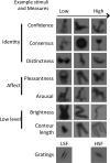
Examples of LSF images and their measures taken along 7 dimensions. Identity measures were confidence, consensus, and distinctness. Affect measures were pleasantness and arousal. Low-level measures were average luminance and contour length. Two exemplar gratings are also shown in the last row.
Figure 2.
Figure 2.
Schematic trial structure of one epoch. Objects and gratings were presented in a mixed pseudorandomized order. Subjects had to press 1 of the 2 buttons in response to each stimulus to indicate whether or not that stimulus was the same as the one presented 2 stimuli before. There were 4–5 such targets per epoch of 16 trials (4 represented here). A resting period of 10 s was presented after each epoch. Thirty-three such epochs were presented to each subject. Only the responses to correct “No” responses to unrepeated stimuli were analyzed. All images were associated with a measure from each of the dimensions presented in Figure 1, which were used as covariates in the general linear model of the data.
Figure 3.
Figure 3.
Response pattern of the confidence reactive region in the medial OFC, and whole-brain response to LSF images compared with gratings. (A) Parametric modulation of image response by confidence in the OFC. The effect of the confidence parametric regressor is shown in red, and the overall response to images versus gratings is shown in blue. The bar graph on the right shows activations within the cluster responding to confidence. They are not independent of the isolated contrast cluster and are shown for illustrating the effect. (B) Greater response to LSF images than to gratings. Response of images with low (blue), medium (cyan), and high (green) degrees of confidence are shown in horizontal slices of the average brain anatomy. Z-coordinate increments in steps of 3 mm from left to right. (C) Bar plots showing the response to images compared with rest periods in 4 different regions indicated above each graph. The Y-coordinates of these regions correspond to the thin dotted white lines overlayed on the maps in B. The black bars represent the response to the gratings and the 3 levels of confidence use the same color code as on the brain slices in B. All coordinates are in the Montreal Neurological Institute frame. For all maps, significance was assessed by means of nonparametric permutation statistics corrected for family-wise error rate (P < 0.05).
Figure 4.
Figure 4.
PPI effects between the lateral OFC region, confidence, and activity in regions more active in response to images than to gratings. Left: significant PPI regions (red) for the lateral OFC ROI (yellow) within the regions more active in response to images than to gratings (blue). Effects are displayed on the average structural brain on 3 orthogonal slices centered at coordinates: x = 48, y = 36, z = −9. Right: Same as left, but displayed on a transparent glassbrain. For all maps, testing was restricted to the regions significantly more active in response to LSF images than to gratings. Significance was assessed by means of nonparametric permutation statistics corrected for family-wise error rate (P < 0.05).

References

    1. Andrade A, Paradis AL, Rouquette S, Poline JB. Ambiguous results in functional neuroimaging data analysis due to covariate correlation. NeuroImage. 1999;10:483–486. - PubMed
    1. Bachevalier J, Mishkin M. Visual recognition impairment follows ventromedial but not dorsolateral prefrontal lesions in monkeys. Behav Brain Res. 1986;20:249–261. - PubMed
    1. Bar M. Visual objects in context. Nat Rev Neurosci. 2004;5:617–629. - PubMed
    1. Bar M, Kassam K, Ghuman A, Boshyan J, Schmid A, Dale A, Häamäaläainen M, Marinkovic K, Schacter D, Rosen BR. Top-down facilitation of visual recognition. Proc Natl Acad Sci. 2006;103:449. - PMC - PubMed
    1. Bar M, Tootell RBH, Schacter DL, Greve DN, Fischl B, Mendola JD, Rosen BR, Dale AM. Cortical mechanisms specific to explicit visual object recognition. Neuron. 2001;29:529–535. - PubMed

Publication types