

Opposing roles of STAT4 and Dnmt3a in Th1 gene regulation
- PMID: 23772023
- PMCID: PMC3703830
- DOI: 10.4049/jimmunol.1203229
Opposing roles of STAT4 and Dnmt3a in Th1 gene regulation
Abstract
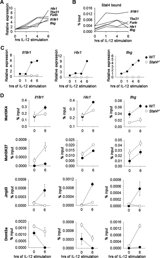
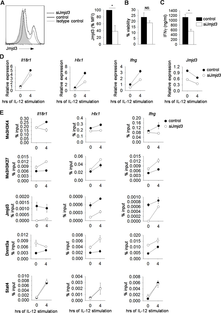
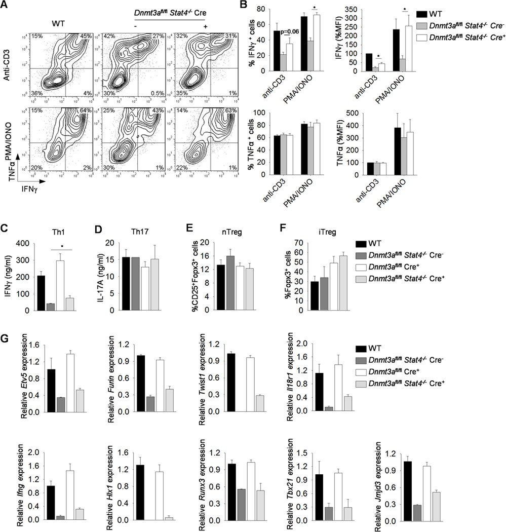
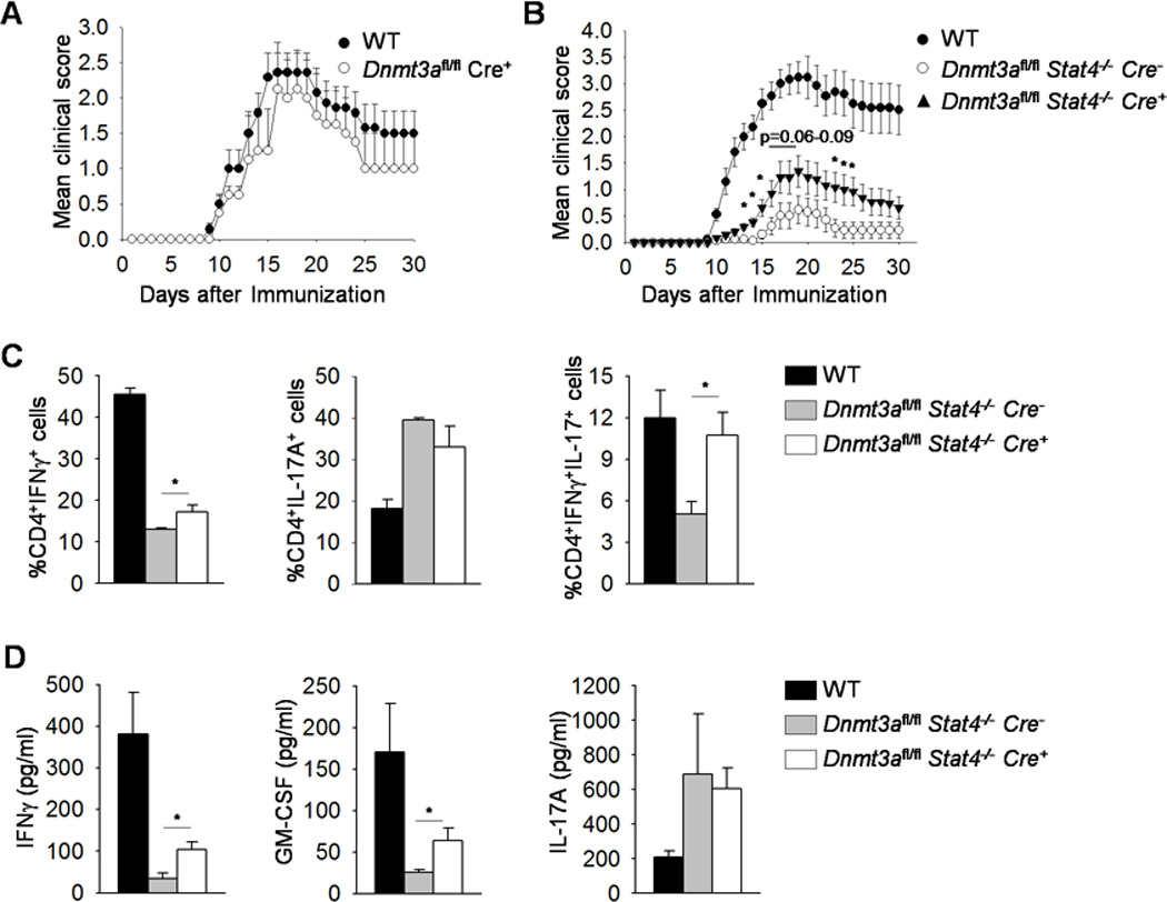
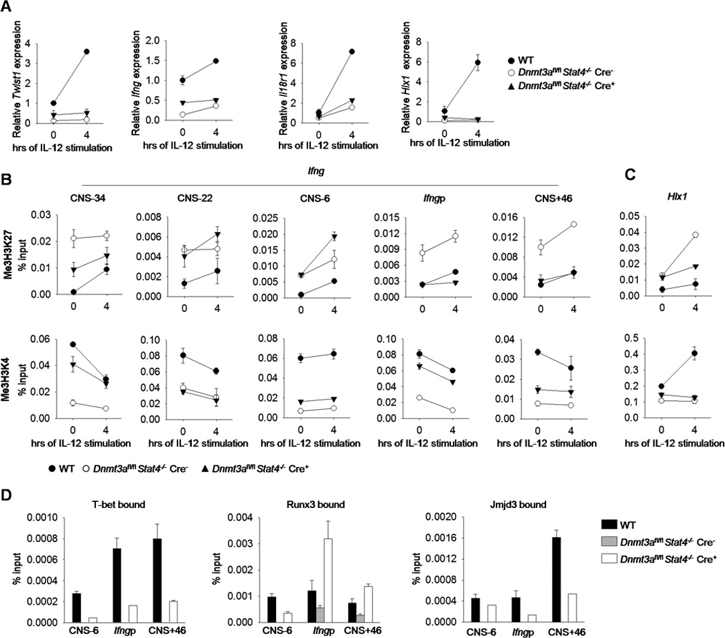
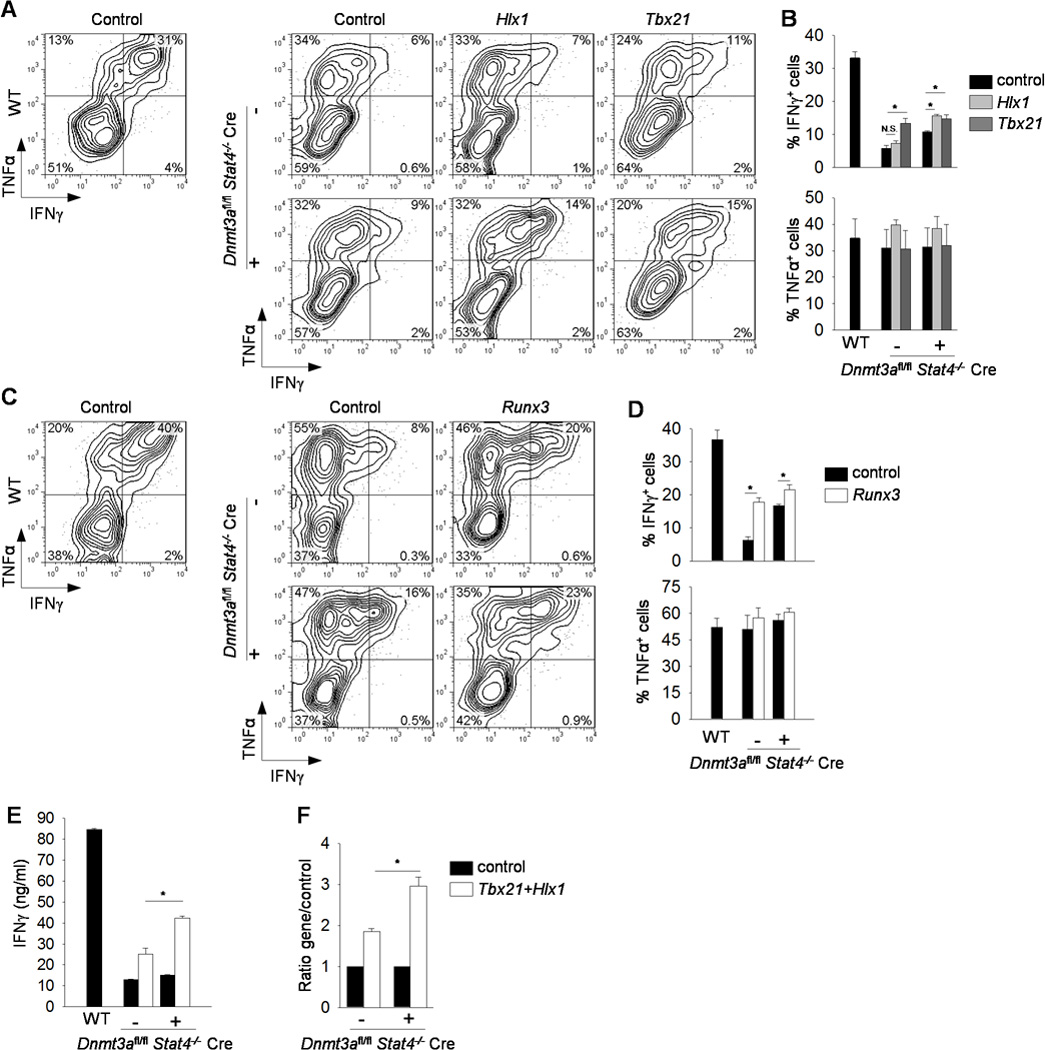
The STAT transcription factor STAT4 is a critical regulator of Th1 differentiation and inflammatory disease. Yet, how STAT4 regulates gene expression is still unclear. In this report, we define a STAT4-dependent sequence of events including histone H3 lysine 4 methylation, Jmjd3 association with STAT4 target loci, and a Jmjd3-dependent decrease in histone H3 lysine 27 trimethylation and DNA methyltransferase (Dnmt) 3a association with STAT4 target loci. Dnmt3a has an obligate role in repressing Th1 gene expression, and in Th1 cultures deficient in both STAT4 and Dnmt3a, there is recovery in the expression of a subset of Th1 genes that is sufficient to increase IFN-γ production. Moreover, although STAT4-deficient mice are protected from the development of experimental autoimmune encephalomyelitis, mice deficient in STAT4 and conditionally deficient in Dnmt3a in T cells develop paralysis. Th1 genes that are derepressed in the absence of Dnmt3a have greater induction after the ectopic expression of the Th1-associated transcription factors T-bet and Hlx1. Together, these data demonstrate that STAT4 and Dnmt3a play opposing roles in regulating Th1 gene expression, and that one mechanism for STAT4-dependent gene programming is in establishing a derepressed genetic state susceptible to transactivation by additional fate-determining transcription factors.
Figures
Similar articles
-
STAT4 controls GM-CSF production by both Th1 and Th17 cells during EAE.J Neuroinflammation. 2015 Jun 30;12:128. doi: 10.1186/s12974-015-0351-3. J Neuroinflammation. 2015. PMID: 26123499 Free PMC article.
-
Stat4 limits DNA methyltransferase recruitment and DNA methylation of the IL-18Ralpha gene during Th1 differentiation.EMBO J. 2007 Apr 18;26(8):2052-60. doi: 10.1038/sj.emboj.7601653. Epub 2007 Mar 22. EMBO J. 2007. PMID: 17380127 Free PMC article.
-
Signal transducer and activator of transcription 4 is required for the transcription factor T-bet to promote T helper 1 cell-fate determination.Immunity. 2008 Nov 14;29(5):679-90. doi: 10.1016/j.immuni.2008.08.017. Immunity. 2008. PMID: 18993086 Free PMC article.
-
Stat4 is critical for the balance between Th17 cells and regulatory T cells in colitis.J Immunol. 2011 Jun 1;186(11):6597-606. doi: 10.4049/jimmunol.1004074. Epub 2011 Apr 27. J Immunol. 2011. PMID: 21525389 Free PMC article.
-
Intestinal irradiation and fibrosis in a Th1-deficient environment.Int J Radiat Oncol Biol Phys. 2012 Sep 1;84(1):266-73. doi: 10.1016/j.ijrobp.2011.11.027. Epub 2012 Feb 13. Int J Radiat Oncol Biol Phys. 2012. PMID: 22336200
Cited by
-
STAT4 controls GM-CSF production by both Th1 and Th17 cells during EAE.J Neuroinflammation. 2015 Jun 30;12:128. doi: 10.1186/s12974-015-0351-3. J Neuroinflammation. 2015. PMID: 26123499 Free PMC article.
-
AP-1 activity induced by co-stimulation is required for chromatin opening during T cell activation.J Exp Med. 2020 Jan 6;217(1):e20182009. doi: 10.1084/jem.20182009. J Exp Med. 2020. PMID: 31653690 Free PMC article.
-
Dnmt3a-dependent de novo DNA methylation enforces lineage commitment and preserves functionality of memory Th1 and Tfh cells.bioRxiv [Preprint]. 2025 Feb 15:2024.12.03.623450. doi: 10.1101/2024.12.03.623450. bioRxiv. 2025. PMID: 39677644 Free PMC article. Preprint.
-
Role of IL-18 induced Amphiregulin expression on virus induced ocular lesions.Mucosal Immunol. 2018 Nov;11(6):1705-1715. doi: 10.1038/s41385-018-0058-8. Epub 2018 Aug 7. Mucosal Immunol. 2018. PMID: 30087443 Free PMC article.
-
Clonal hematopoiesis, somatic mosaicism, and age-associated disease.Physiol Rev. 2023 Jan 1;103(1):649-716. doi: 10.1152/physrev.00004.2022. Epub 2022 Sep 1. Physiol Rev. 2023. PMID: 36049115 Free PMC article. Review.
References
-
- Kaplan MH. STAT4: A critical regulator of inflammation in vivo. Immunologic Research. 2005;32:231–241. - PubMed
-
- Watford WT, Hissong BD, Bream JH, Kanno Y, Muul L, O'Shea JJ. Signaling by IL-12 and IL-23 and the immunoregulatory roles of STAT4. Immunol Rev. 2004;202:139–156. - PubMed
-
- Hsieh C-S, Macatonia SE, Tripp CS, Wolf SF, O'Garra A, Murphy KM. Development of Th1 CD4+ T cells through IL-12 produced by Listeria-induced macrophages. Science. 1993;260:547–549. - PubMed
-
- Afkarian M, Sedy JR, Yang J, Jacobson NG, Cereb N, Yang SY, Murphy TL, Murphy KM. T-bet is a STAT1-induced regulator of IL-12R expression in naive CD4+ T cells. Nat Immunol. 2002;3:549–557. - PubMed
-
- Wenner CA, Güler ML, Macatonia SE, O'Garra A, Murphy KM. Roles of IFN-γ and IFN-α in IL-12-induced T helper cell-1 development. J Immunol. 1996;156:1442–1447. - PubMed
Publication types
MeSH terms
Substances
Grants and funding
LinkOut - more resources
Full Text Sources
Other Literature Sources
Molecular Biology Databases
Miscellaneous
