

Cutting edge: Conditional MHC class II expression reveals a limited role for B cell antigen presentation in primary and secondary CD4 T cell responses
- PMID: 23772037
- PMCID: PMC3711531
- DOI: 10.4049/jimmunol.1201598
Cutting edge: Conditional MHC class II expression reveals a limited role for B cell antigen presentation in primary and secondary CD4 T cell responses
Abstract
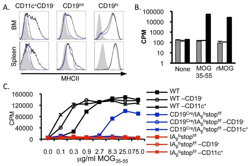
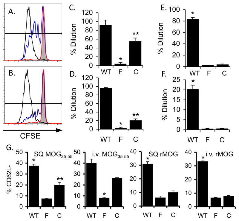
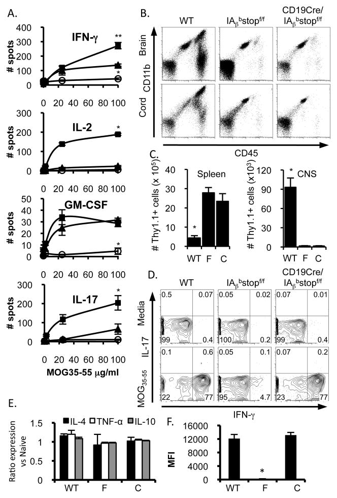
The activation, differentiation, and subsequent effector functions of CD4 T cells depend on interactions with a multitude of MHC class II (MHCII)-expressing APCs. To evaluate the individual contribution of various APCs to CD4 T cell function, we have designed a new murine tool for selective in vivo expression of MHCII in subsets of APCs. Conditional expression of MHCII in B cells was achieved using a cre-loxP approach. After i.v. or s.c. priming, partial proliferation and activation of CD4 T cells was observed in mice expressing MHCII only by B cells. Restricting MHCII expression to B cells constrained secondary CD4 T cell responses in vivo, as demonstrated in a CD4 T cell-dependent model of autoimmunity, experimental autoimmune encephalomyelitis. These results highlight the limitations of B cell Ag presentation during initiation and propagation of CD4 T cell function in vivo using a novel system to study individual APCs by the conditional expression of MHCII.
Figures
Similar articles
-
MHC class II-restricted antigen presentation by plasmacytoid dendritic cells inhibits T cell-mediated autoimmunity.J Exp Med. 2010 Aug 30;207(9):1891-905. doi: 10.1084/jem.20092627. Epub 2010 Aug 9. J Exp Med. 2010. PMID: 20696698 Free PMC article.
-
CD8alpha+ and CD11b+ dendritic cell-restricted MHC class II controls Th1 CD4+ T cell immunity.J Immunol. 2003 Nov 15;171(10):5077-84. doi: 10.4049/jimmunol.171.10.5077. J Immunol. 2003. PMID: 14607905
-
Modified vaccinia virus Ankara-infected dendritic cells present CD4+ T-cell epitopes by endogenous major histocompatibility complex class II presentation pathways.J Virol. 2015 Mar;89(5):2698-709. doi: 10.1128/JVI.03244-14. Epub 2014 Dec 17. J Virol. 2015. PMID: 25520512 Free PMC article.
-
The role of B cell antigen presentation in the initiation of CD4+ T cell response.Immunol Rev. 2020 Jul;296(1):24-35. doi: 10.1111/imr.12859. Epub 2020 Apr 18. Immunol Rev. 2020. PMID: 32304104 Review.
-
How Does B Cell Antigen Presentation Affect Memory CD4 T Cell Differentiation and Longevity?Front Immunol. 2021 Jun 10;12:677036. doi: 10.3389/fimmu.2021.677036. eCollection 2021. Front Immunol. 2021. PMID: 34177919 Free PMC article. Review.
Cited by
-
Establishment of fetomaternal tolerance through glycan-mediated B cell suppression.Nature. 2022 Mar;603(7901):497-502. doi: 10.1038/s41586-022-04471-0. Epub 2022 Mar 2. Nature. 2022. PMID: 35236989 Free PMC article.
-
MicroRNA signature of central nervous system-infiltrating dendritic cells in an animal model of multiple sclerosis.Immunology. 2018 Sep;155(1):112-122. doi: 10.1111/imm.12934. Epub 2018 May 10. Immunology. 2018. PMID: 29749614 Free PMC article.
-
B Cells Influence Encephalitogenic T Cell Frequency to Myelin Oligodendrocyte Glycoprotein (MOG)38-49 during Full-length MOG Protein-Induced Demyelinating Disease.Immunohorizons. 2024 Sep 1;8(9):729-739. doi: 10.4049/immunohorizons.2400069. Immunohorizons. 2024. PMID: 39330967 Free PMC article.
-
B cell antigen presentation in the initiation of follicular helper T cell and germinal center differentiation.J Immunol. 2014 Apr 15;192(8):3607-17. doi: 10.4049/jimmunol.1301284. Epub 2014 Mar 19. J Immunol. 2014. PMID: 24646739 Free PMC article.
-
cDC1 prime and are licensed by CD4+ T cells to induce anti-tumour immunity.Nature. 2020 Aug;584(7822):624-629. doi: 10.1038/s41586-020-2611-3. Epub 2020 Aug 12. Nature. 2020. PMID: 32788723 Free PMC article.
References
-
- Reis e Sousa C. Dendritic cells in a mature age. Nat Rev Immunol. 2006;6:476–483. - PubMed
-
- Pape KA, Catron DM, Itano AA, Jenkins MK. The humoral immune response is initiated in lymph nodes by B cells that acquire soluble antigen directly in the follicles. Immunity. 2007;26:491–502. - PubMed
-
- Banchereau J, Steinman RM. Dendritic cells and the control of immunity. Nature. 1998;392:245–252. - PubMed
-
- Constant SL. B lymphocytes as antigen-presenting cells for CD4+ T cell priming in vivo. J Immunol. 1999;162:5695–5703. - PubMed
Publication types
MeSH terms
Substances
Grants and funding
LinkOut - more resources
Full Text Sources
Other Literature Sources
Molecular Biology Databases
Research Materials
Miscellaneous
