Long-term continuous positive airway pressure therapy normalizes high exhaled nitric oxide levels in obstructive sleep apnea
- PMID: 23772184
- PMCID: PMC3659371
- DOI: 10.5664/jcsm.2740
Long-term continuous positive airway pressure therapy normalizes high exhaled nitric oxide levels in obstructive sleep apnea
Abstract
Study objectives: Upper airway inflammation and oxidative stress have been implicated in the pathogenesis of obstructive sleep apnea (OSA) and may be linked to cardiovascular consequences. We prospectively examined fraction of exhaled nitric oxide (FENO), a surrogate marker of upper airway inflammation using a portable nitric oxide analyzer (NIOX MINO).
Design: In consecutive adult nonsmokers with suspected OSA, FENO was measured immediately before and after polysomnographic studies, and within 1-3 months following continuous positive airway pressure (CPAP) therapy.
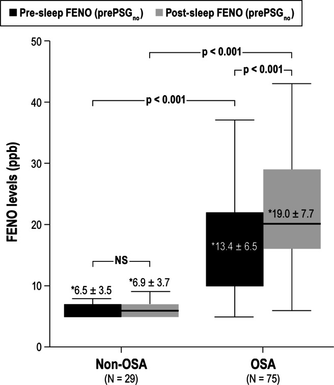
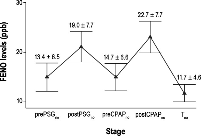
Measurement and results: FENO levels were increased in the 75 patients with OSA compared to the 29 controls, both before sleep (13.4 ± 6.5 ppb vs. 6.5 ± 3.5; p < 0.001) and after sleep (19.0 ± 7.7 ppb vs. 6.9 ± 3.7; p < 0.001). Furthermore, in patients with OSA, FENO levels were significantly higher post-sleep than pre-sleep (19.0 ± 7.7 ppb vs. 13.4 ± 6.5; p < 0.001), while there was no significant overnight change in patients without OSA. The rise in FENO correlated with the apnea-hypopnea index (r = 0.65, p < 0.001), nadir oxygen saturation (r = 0.54, p < 0.001), and arousal index (r = 0.52, p < 0.001). Thirty-seven of these patients underwent CPAP titration and treatment. Successful titration was associated with a lower overnight increase in FENO (7.2 ± 3.3 vs. 11.0 ± 4.3, p = 0.02). FENO levels declined after 1-3 months of CPAP therapy (11.7 ± 4.4 ppb, p < 0.001).
Conclusions: FENO levels are elevated in OSA, correlate with severity, and decrease after positive pressure therapy. This study supports the role of upper airway inflammation in OSA pathogenesis and a possible role for FENO in monitoring CPAP therapy.
Keywords: Sleep disordered breathing; endogenous nitrate vasodilator; positive pressure ventilation.
Figures


Comment in
-
High-dose N-acetylcysteine in chronic obstructive pulmonary disease, prone positioning in acute respiratory distress syndrome, and continuous positive airway pressure and exhaled nitric oxide in obstructive sleep apnea.Am J Respir Crit Care Med. 2014 Jan 15;189(2):223-4. doi: 10.1164/rccm.201308-1555RR. Am J Respir Crit Care Med. 2014. PMID: 24428648 Free PMC article. No abstract available.
Similar articles
-
Measurement of fractional exhaled nitric oxide and nasal nitric oxide in male patients with obstructive sleep apnea.Sleep Breath. 2019 Sep;23(3):785-793. doi: 10.1007/s11325-018-1760-1. Epub 2018 Dec 12. Sleep Breath. 2019. PMID: 30542936 Free PMC article.
-
Nasal nitric oxide improved by continuous positive airway pressure therapy for upper airway inflammation in obstructive sleep apnea.Sleep Breath. 2017 May;21(2):405-410. doi: 10.1007/s11325-016-1431-z. Epub 2016 Nov 12. Sleep Breath. 2017. PMID: 27837378
-
Effect of Continuous Positive Airway Pressure on Airway Inflammation and Oxidative Stress in Patients with Obstructive Sleep Apnea.Can Respir J. 2016;2016:3107324. doi: 10.1155/2016/3107324. Epub 2016 Jun 30. Can Respir J. 2016. PMID: 27445526 Free PMC article.
-
Impact of continuous positive airway pressure on vascular endothelial growth factor in patients with obstructive sleep apnea: a meta-analysis.Sleep Breath. 2019 Mar;23(1):5-12. doi: 10.1007/s11325-018-1660-4. Epub 2018 Apr 18. Sleep Breath. 2019. PMID: 29671205 Review.
-
The Role of Aldosterone in OSA and OSA-Related Hypertension.Front Endocrinol (Lausanne). 2022 Jan 12;12:801689. doi: 10.3389/fendo.2021.801689. eCollection 2021. Front Endocrinol (Lausanne). 2022. PMID: 35095768 Free PMC article. Review.
Cited by
-
Impact of Asthma and Obstructive Sleep Apnea on Central Airways Resistance During Pregnancy.Lung. 2025 Jun 3;203(1):66. doi: 10.1007/s00408-025-00817-3. Lung. 2025. PMID: 40459639
-
Sleep-disordered breathing children: Measurement of nasal nitric oxide and fractional exhaled nitric oxide.HNO. 2016 Mar;64(3):169-74. doi: 10.1007/s00106-016-0120-3. HNO. 2016. PMID: 26952133
-
Gestational Obstructive Sleep Apnea: Biomarker Screening Models and Lack of Postpartum Resolution.J Clin Sleep Med. 2018 Apr 15;14(4):549-555. doi: 10.5664/jcsm.7042. J Clin Sleep Med. 2018. PMID: 29609706 Free PMC article.
-
Study of Exhaled Nitric Oxide in Subjects with Suspected Obstructive Sleep Apnea: A Pilot Study in Vietnam.Pulm Med. 2016;2016:3050918. doi: 10.1155/2016/3050918. Epub 2016 Jan 13. Pulm Med. 2016. PMID: 26881073 Free PMC article. Clinical Trial.
-
Psoriasis and Respiratory Comorbidities: The Added Value of Fraction of Exhaled Nitric Oxide as a New Method to Detect, Evaluate, and Monitor Psoriatic Systemic Involvement and Therapeutic Efficacy.Biomed Res Int. 2018 Sep 23;2018:3140682. doi: 10.1155/2018/3140682. eCollection 2018. Biomed Res Int. 2018. PMID: 30345297 Free PMC article. Review.
References
-
- Berger G, Gilbey P, Hammel I, Ophir D. Histopathology of the uvula and the soft palate in patients with mild, moderate, and severe obstructive sleep apnea. Laryngoscope. 2002;112:357–63. - PubMed
-
- Paulsen FP, Steven P, Tsokos M, et al. Upper airway epithelial structural changes in obstructive sleep-disordered breathing. Am J Respir Crit Care Med. 2002;166:501–9. - PubMed
-
- Zakkar M, Sekosan M, Wenig B, Olopade CO, Rubinstein I. Decrease in immunoreactive neutral endopeptidase in uvula epithelium of patients with obstructive sleep apnea. Ann Otol Rhinol Laryngol. 1997;106:474–7. - PubMed
-
- Sekosan M, Zakkar M, Wenig BL, Olopade CO, Rubinstein I. Inflammation in the uvula mucosa of patients with obstructive sleep apnea. Laryngoscope. 1996;106:1018–20. - PubMed
-
- Kharitonov SA, Barnes PJ. Exhaled markers of pulmonary disease. Am J Respir Crit Care Med. 2001;163:1693–722. - PubMed
Publication types
MeSH terms
Substances
Grants and funding
LinkOut - more resources
Full Text Sources
Other Literature Sources