. 2013 Jun 10:7:86.
doi: 10.3389/fncel.2013.00086. eCollection 2013.

Sonic hedgehog and Wnt: antagonists in morphogenesis but collaborators in axon guidance

Affiliations

Sonic hedgehog and Wnt: antagonists in morphogenesis but collaborators in axon guidance

Evelyn C Avilés et al. Front Cell Neurosci. .

Abstract

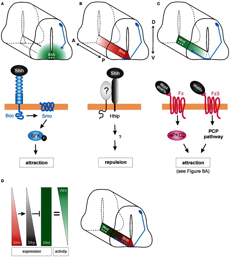
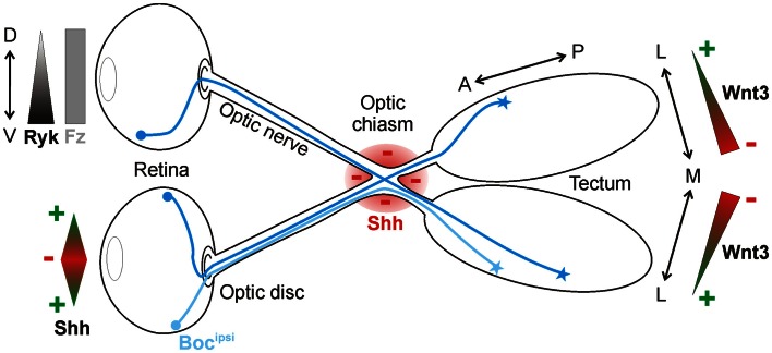
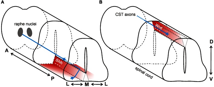
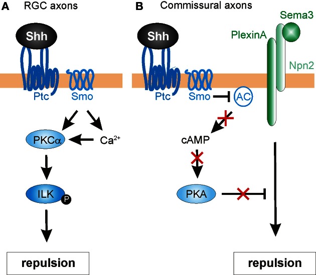
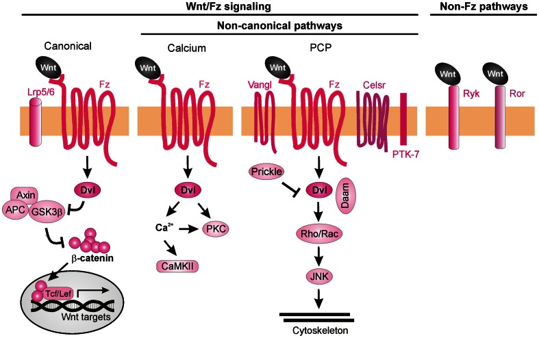
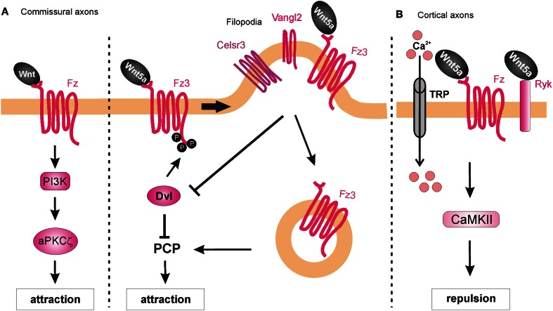
As indicated by their name, morphogens were first identified for their role in the formation of tissues early in development. Secreted from a source, they spread through the tissue to form gradients by which they affect the differentiation of precursor cells in a concentration-dependent manner. In this context, the antagonistic roles of the morphogens of the Wnt family and Sonic hedgehog (Shh) in the specification of cell types along the dorso-ventral axis of the neural tube have been studied in detail. However, more recently, morphogens have been demonstrated to act well beyond the early stages of nervous system development, as additional roles of morphogen gradients in vertebrate neural circuit formation have been identified. Both Wnt and Shh affect neural circuit formation at several stages by their influence on neurite extension, axon pathfinding and synapse formation. In this review, we will summarize the mechanisms of morphogen function during axon guidance in the vertebrate nervous system.

Keywords: Frizzled; Ryk; Smoothened; attraction; morphogen; neural circuit; repulsion; spinal cord.


Figures

Figure 1
Figure 1
Antagonistic activities of Shh and Wnt/BMP pattern the developing spinal cord. Counteracting gradients of Shh, secreted from the floorplate (FP), and Wnt/BMP, derived from the roof plate (RP) induce the concentration-dependent differentiation of precursor cells along the dorso-ventral (D-V) axis. During morphogenesis, Shh and Wnts have antagonistic functions. Shh promotes the formation of Gli activator forms, while Wnts directly induce the expression of Gli3, which acts as a transcriptional repressor in the absence of Shh (see Dessaud et al., , for details). In turn, the specific combinations of transcription factors induced by Shh and Wnts generate a cell identity code that specifies the neural progenitor subtypes. As these cells exit the cell cycle, they distribute laterally in a specific order along the dorso-ventral axis (dI1-v3).
Figure 2
Figure 2
Shh and Wnts guide commissural axons in the vertebrate spinal cord. (A) Pre-crossing commissural axons (blue) are attracted ventrally toward the midline by an increasing gradient of Shh produced in the floorplate (green). The attractive effect of Shh is mediated by Smoothened (Smo) and Brother of CDO (Boc) in a transcription-independent manner. Instead, the activation of Src family kinases (SFK) induces cytoskeletal rearrangements in the growth cone. (B) The response of commissural axons to Shh switches from attraction to repulsion when axons reach the midline. Post-crossing commissural axons are pushed anteriorly by a posteriorhigh to anteriorlow gradient of Shh (red). The repellent activity of Shh is mediated by Hedgehog-interacting protein (Hhip), a receptor that is transiently upregulated on commissural axons at the time of their turning into the longitudinal axis. An additional signaling co-receptor may also be involved. (C) An anteriorhigh to posteriorlow gradient of Wnt activity works in parallel to Shh repulsion to attract post-crossing commissural axons anteriorly. Depending on the species, Wnt4, Wnt5a, and Wnt7a are attractants for post-crossing commissural axons via non-canonical pathways. In mouse, Fz3, in response to Wnt4/Wnt7b, activates a complex containing an atypical protein kinase C (aPKCζ). In response to Wnt5a, the PCP pathway is activated. See text for more details. (D) In chick, Shh was shown to shape Wnt activity indirectly. Wnt5a and Wnt7a are expressed uniformly along the longitudinal axis. In addition to its direct effect on post-crossing commissural axons, Shh induces the expression of the Wnt antagonist Sfrp1 in a posteriorhigh to anteriorlow gradient in the floorplate. The antagonistic activity of Sfrp in turn regulates the activity of Wnts in the floorplate, such that an attractive “activity gradient” of Wnt is formed. Thus, Shh (red) and Wnt (green) gradients collaborate to guide post-crossing commissural axons anteriorly.
Figure 3
Figure 3
Shh and Wnt3 are axon guidance cues in the visual system. In the retina, Shh is bifunctional: low concentrations promote RGC axon outgrowth toward the optic disc, whereas high concentrations push axons into the optic nerve. Shh is also expressed along the border of the optic chiasm (red), where it acts as a repellant for RGC axons in the optic nerve. Ipsilaterally projecting RGC axons express Boc (Bocipsi), which mediates Shh repulsion at the chiasm. Wnt3 is expressed in a medialhigh-to-laterallow gradient in the tectum. At high concentrations, Wnt3 inhibits the growth of both dorsal and ventral RGC axons via Ryk as a receptor. At low concentrations, Wnt3 stimulates the growth of dorsal RGC axons to the lateral tectum in a Fz-dependent manner.
Figure 4
Figure 4
Wnt and Shh are repellants for descending axon tracts of the brain. (A) In mouse, Shh repels axons of the raphe spinal tract (blue) multidirectionally. In the anterior spinal cord, Shh (red) is expressed in an anteriorhigh to posteriorlow (A-P) gradient, and in a medialhigh to laterallow (M-L) gradient. Thus, RST axons are pushed to grow both posteriorly and laterally. (B) In mouse, a decreasing anteriorhigh to posteriorlow Wnt gradient (red) in the roofplate of the dorsal spinal cord repels descending corticospinal tract axons (blue) posteriorly.
Figure 5
Figure 5
Intracellular mechanisms mediating Shh-induced repulsive axon guidance. (A) In RGC axons, Shh binding to Ptc promotes the Smo-dependent activation of protein kinase Cα (PKCα), which in turn phosphorylates integrin-linked kinase (ILK). ILK promotes repulsive axon turning. (B) In commissural neurons, Shh acts through Ptc and Smo to block adenylyl cyclase (AC) activity. This lowers cAMP levels and inhibits protein kinase A (PKA) activity. In turn, this confers growth cone sensitivity to class 3 semaphorins (Sema3), by allowing repulsive signaling downstream of a PlexinA-Npn2 complex.
Figure 6
Figure 6
The canonical Shh signaling pathway. In the absence of Shh, Gli transcription factors are proteolytically processed to repressor forms (GliR), which block the transcription of target genes. Shh binding to the 12-pass transmembrane receptor Patched (Ptc) relieves the inhibition of Smoothened (Smo), which in turn promotes the accumulation of the activator forms of Gli (GliA) while suppressing GliR production. The regulation of Gli activity occurs via an intracellular complex composed of Fused (Fu), Suppressor of Fused (SuFu) and possibly other components (?).
Figure 7
Figure 7
Wnt signaling pathways. Wnt ligands can transduce their signal through at least three Frizzled-dependent (Fz) pathways: the canonical, the calcium, and the PCP pathways, all of which involve the activation of Dishevelled (Dvl). The canonical pathway requires the co-receptors Lrp5/6 to recruit Dvl and inhibit the “destruction complex” composed of Adenomatous polyposis coli (APC), Axin, and Glycogen synthase 3β (GSK3β). Wnt binding leads to the phosphorylation of GSK3β in this complex and, as a consequence, to the accumulation of unphosphorylated β-catenin, which can enter the nucleus and together with the transcription factors Tcf/Lef induce the expression of target genes. Activation of the calcium pathway results in an increase of cytosolic calcium (Ca2+) and the subsequent activation of calcium-dependent kinases (CaMKII). In the PCP pathway, activation of the transmembrane proteins Vangl, Celsr, and PTK7 and the recruitment of the intracellular proteins Prickle and Daam lead to activation of Rho GTPases and JNK, promoting cytoskeleton remodeling. More recently, Fz-independent Wnt signaling has been described. Wnt ligands can bind directly to receptors such as Ryk or Ror. The intracellular signaling cascades activated in these cases are poorly understood.
Figure 8
Figure 8
Intracellular mechanisms underlying Wnt-induced axon guidance. (A) Wnts promote commissural axon attraction via the activation of the PI3K-atypical PKCζ signaling and the PCP pathway. The PCP pathway is inhibited through the Dvl-dependent phosphorylation of Fz3, which as a result remains in the plasma membrane. The local expression of Vangl2 in the filopodia of the growth cone prevents the Dvl-induced phosphorylation of Fz3 upon binding of Wnt5a and results in the internalization of Fzd3 and the activation of the PCP pathway. (B) Wnt5a repels cortical axons by binding to both Fz and Ryk receptors, which allow the entrance of calcium via the TRP channels. Activation of Ryk alone results in axon growth promotion, whereas co-activation of Ryk and Fz is required for axon repulsion.

References

    1. Andre P., Wang Q., Wang N., Gao B., Schilit A., Halford M. M., et al. (2012). The Wnt coreceptor Ryk regulates Wnt/planar cell polarity by modulating the degradation of the core planar cell polarity component Vangl2. J. Biol. Chem. 287, 44518–44525 10.1074/jbc.M112.414441 - DOI - PMC - PubMed
    1. Bagri A., Marin O., Plump A. S., Mak J., Pleasure S. J., Rubenstein J. L., et al. (2002). Slit proteins prevent midline crossing and determine the dorsoventral position of major axonal pathways in the mammalian forebrain. Neuron 33, 233–248 10.1016/S0896-6273(02)00561-5 - DOI - PubMed
    1. Barresi M. J. F., Hutson L. D., Chien C., Karlstrom R. O. (2005). Hedgehog regulated Slit expression determines commissure and glial cell position in the zebrafish forebrain. Development 132, 3643–3656 10.1242/dev.01929 - DOI - PubMed
    1. Birgbauer E., Cowan C. A., Sretavan D. W., Henkemeyer M. (2000). Kinase independent function of EphB receptors in retinal axon pathfinding to the optic disc from dorsal but not ventral retina. Development 127, 1231–1241 - PubMed
    1. Blaess S., Corrales J. D., Joyner A. L. (2006). Sonic hedgehog regulates Gli activator and repressor functions with spatial and temporal precision in the mid/hindbrain region. Development 133, 1799–1809 10.1242/dev.02339 - DOI - PubMed