. 2013 Jun 10:4:138.
doi: 10.3389/fimmu.2013.00138. eCollection 2013.

The dendritic cell response to classic, emerging, and homeostatic danger signals. Implications for autoimmunity

Affiliations

The dendritic cell response to classic, emerging, and homeostatic danger signals. Implications for autoimmunity

Paul M Gallo et al. Front Immunol. .

Abstract

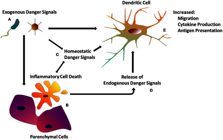
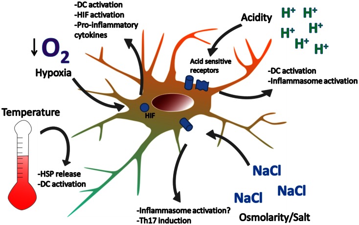
Dendritic cells (DCs) initiate and control immune responses, participate in the maintenance of immunological tolerance and are pivotal players in the pathogenesis of autoimmunity. In patients with autoimmune disease and in experimental animal models of autoimmunity, DCs show abnormalities in both numbers and activation state, expressing immunogenic levels of costimulatory molecules and pro-inflammatory cytokines. Exogenous and endogenous danger signals activate DCs to stimulate the immune response. Classic endogenous danger signals are released, activated, or secreted by host cells and tissues experiencing stress, damage, and non-physiologic cell death; and are therefore referred to as damage-associated molecular patterns (DAMPs). Some DAMPs are released from cells, where they are normally sequestered, during necrosis (e.g., heat shock proteins, uric acid, ATP, HMGB1, mitochondria-derived molecules). Others are actively secreted, like Type I Interferons. Here we discuss important DAMPs in the context of autoimmunity. For some, there is a clear pathogenic link (e.g., nucleic acids and lupus). For others, there is less evidence. Additionally, we explore emerging danger signals. These include inorganic materials and man-made technologies (e.g., nanomaterials) developed as novel therapeutic approaches. Some nanomaterials can activate DCs and may trigger unintended inflammatory responses. Finally, we will review "homeostatic danger signals," danger signals that do not derive directly from pathogens or dying cells but are associated with perturbations of tissue/cell homeostasis and may signal pathological stress. These signals, like acidosis, hypoxia, and changes in osmolarity, also play a role in inflammation and autoimmunity.

Keywords: DAMPs; acidosis; autoimmunity; dendritic cells; hypoxia; mitochondria; nanomaterial; osmolarity.


Figures

Figure 1
Figure 1
The dendritic cell response to danger. (A) Exogenous danger signals include pathogen-associated molecular patterns, as well as exogenous particulate matter. Exogenous danger signals activate DCs directly via pattern recognition receptors, or indirectly though tissue damage (B) and homeostatic perturbations (C). (B) Inflammatory cell death as a result of tissue injury or programed necrosis causes the release of endogenous danger signals (D) (see Table 1) which also activate DCs (see Table 1). (C) Homeostatic perturbations such as those found in inflamed tissue (e.g., decreased pH, hypoxia) may also act as endogenous danger signals, influencing DC immune functions. (E) DCs integrate both exogenous and endogenous danger signals in order to orchestrate the appropriate immune response.
Figure 2
Figure 2
Examples of homeostatic danger signals. Localized perturbations in homeostasis are common in infection, ischemia-reperfusion injury, and inflammation associated with autoimmune disease. Extreme temperatures can cause DC activation and cell death. Heat induces the release of HSPs which can activate DC. Hypoxia is sensed by HIF and leads to DC activation, NF-kB signaling, and cytokine production. Acidity is sensed by acid-sensing ion channels and acid-sensing G-proteins, leading to DC activation. Hypotonicity activates the inflammasome in macrophages and high salt concentrations promote Th17 responses. How DCs respond to changes in osmolarity remains unclear.

References

    1. Abdulahad D. A., Westra J., Bijzet J., Limburg P. C., Kallenberg C. G., Bijl M. (2011). High mobility group box 1 (HMGB1) and anti-HMGB1 antibodies and their relation to disease characteristics in systemic lupus erythematosus. Arthritis Res. Ther. 13, R71.10.1186/ar3332 - DOI - PMC - PubMed
    1. Adachi Y., Taketani S., Toki J., Ikebukuro K., Sugiura K., Oyaizu H., et al. (2002). Marked increase in number of dendritic cells in autoimmune-prone (NZW x BXSB)F1 mice with age. Stem Cells 20, 61–7210.1634/stemcells.20-1-61 - DOI - PubMed
    1. Aggarwal P., Hall J. B., McLeland C. B., Dobrovolskaia M. A., Mcneil S. E. (2009). Nanoparticle interaction with plasma proteins as it relates to particle biodistribution, biocompatibility and therapeutic efficacy. Adv. Drug Deliv. Rev. 61, 428–43710.1016/j.addr.2009.03.009 - DOI - PMC - PubMed
    1. Amodio G., Gregori S. (2012). Dendritic cells a double-edge sword in autoimmune responses. Front. Immunol. 3:23310.3389/fimmu.2012.00233 - DOI - PMC - PubMed
    1. Andersson A., Covacu R., Sunnemark D., Danilov A., Dal Bianco A., Khademi M., et al. (2008). Pivotal advance: HMGB1 expression in active lesions of human and experimental multiple sclerosis. J. Leukoc. Biol. 84, 1248–125510.1189/jlb.1207844 - DOI - PubMed

LinkOut - more resources