Review
. 2013 Jul;254(1):65-77.
doi: 10.1111/imr.12070.

Pathobiology of HIV/SIV-associated changes in secondary lymphoid tissues

Affiliations
Review

Pathobiology of HIV/SIV-associated changes in secondary lymphoid tissues

Jacob D Estes. Immunol Rev. 2013 Jul.

Abstract

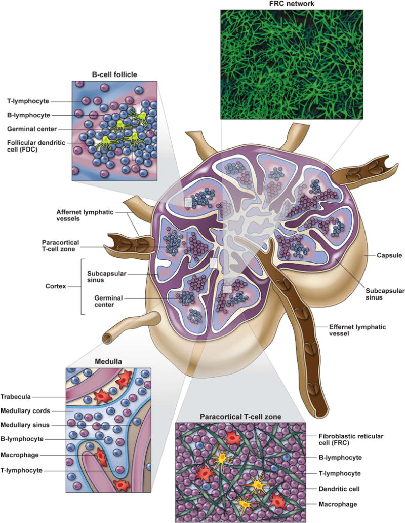
Acquired immunodeficiency syndrome (AIDS) is principally a disease of lymphoid tissues (LTs), due to the fact that the main target cell of human immunodeficiency virus (HIV) is the CD4(+) T lymphocyte that primarily resides within organs of the immune system. The impact of HIV infection on secondary LTs, in particular lymph nodes, is critical to delineate, as these immune organs are the principal sites for initiating and facilitating immune responses and are critical for lymphocyte homeostatic maintenance and survival. The underlying structural elements of LTs, fibroblastic reticular cell (FRC) network, not only form the architectural framework for these organs, but also play in integral role in the production and storage of cytokines needed for T-cell survival. There is an interdependent relationship between the FRC stromal network and CD4(+) T lymphocytes for their survival and maintenance that is progressively disrupted during HIV disease. HIV infection results in profound pathological changes to LTs induced by persistent chronic immune activation and inflammation that leads to progressive collagen deposition and fibrosis disrupting and damaging the important FRC network. In this review, I focus on the process, mechanisms, and the implications of pathological damage to important secondary LTs, combining what we have learned from HIV-infected individuals as well as the invaluable knowledge gained from studies in non-human primate simian immunodeficiency virus infection models.


Figures

Figure 1
Figure 1. Lymph node anatomy and function
LNs are highly organized lymphoid tissues that facilitate immune responses through the orchestrated movement and interactions of lymphocytes with their respective antigen presenting cell (APC) populations (i.e. DCs for T cells and FDCs for B cells). Lymphocytes and APCs enter the LN either through the afferent lymphatic vessels or HEVs and migrate along the FRC network in response to chemotactic cues. Activated B cells primarily migrate to the superficial cortical region of LNs where B cell follicles are abundantly located. B cells entering follicles rapidly proliferate to form germinal centers and interact with FDCs and TFH to mount humoral immune responses. Most T lymphocytes (except specialized TFH that migrate to B cell follicles) transvers to the paracortical T cell zone (TZ) located intermediate to the superficial cortex and deep medullary cords. Within the TZ, T cells continually migrate along the FRC network interacting with numerous DCs interspersed throughout this 3-deminsional meshwork, sampling MHC-peptide complexes until their TCR binds to their cognate MHC-peptide match at which time they stop migrating, receive activation signals and undergo clonal expansion. FRCs are an important source of homeostatic cytokines, especially IL-7, that are needed for T cell survival during their transition through LTs. Lymphocytes that do not bind to their cognate antigen, egress from LNs through the medullary cords and efferent lymphatic vessel. Lymph fluid contains abundant particulate antigens that need to be removed before it can be recirculated through the blood. LNs serve as an important filtration system for bodily fluids, and this lymph filtration primarily occurs through the medullary
Figure 2
Figure 2. Progressive pathological lymph node fibrosis in HIV/SIV infection
Cartoon schematic (top panel) showing the effect of progressive fibrosis on CD4+ T cell and FRC loss during different stages of HIV/SIV infection. NHP SIV-infection models have provided important insight into the pathological damage of LTs at different stages of disease, with particular insight into the acute phase. Progressive collagen (middle panel; brown IHC staining) and fibronectin (bottom panel; red immunefluorescent staining) deposition occurs early during the acute phase and continues through the chronic phase of SIV infection as a consequence of persistent chronic immune activation and inflammation.

References

    1. WHO. http://www.who.int/mediacentre/factsheets/fs360/en/index.html.2012.
    1. Di Mascio M, et al. Noninvasive in vivo imaging of CD4 cells in simian-human immunodeficiency virus (SHIV)-infected nonhuman primates. Blood. 2009;114:328–337. - PMC - PubMed
    1. Trepel F. Number and distribution of lymphocytes in man. A critical analysis. Klin Wochenschr. 1974;52:511–515. - PubMed
    1. Butcher EC, Picker LJ. Lymphocyte homing and homeostasis. Science. 1996;272:60–66. - PubMed
    1. Willard-Mack CL. Normal structure, function, and histology of lymph nodes. Toxicol Pathol. 2006;34:409–424. - PubMed

Publication types

MeSH terms