Review
. 2013 Jul;254(1):143-69.
doi: 10.1111/imr.12074.

Immunology of pediatric HIV infection

Affiliations
Review

Immunology of pediatric HIV infection

Nicole H Tobin et al. Immunol Rev. 2013 Jul.

Abstract

Most infants born to human immunodeficiency virus (HIV)-infected women escape HIV infection. Infants evade infection despite an immature immune system and, in the case of breastfeeding, prolonged repetitive exposure. If infants become infected, the course of their infection and response to treatment differs dramatically depending upon the timing (in utero, intrapartum, or during breastfeeding) and potentially the route of their infection. Perinatally acquired HIV infection occurs during a critical window of immune development. HIV's perturbation of this dynamic process may account for the striking age-dependent differences in HIV disease progression. HIV infection also profoundly disrupts the maternal immune system upon which infants rely for protection and immune instruction. Therefore, it is not surprising that infants who escape HIV infection still suffer adverse effects. In this review, we highlight the unique aspects of pediatric HIV transmission and pathogenesis with a focus on mechanisms by which HIV infection during immune ontogeny may allow discovery of key elements for protection and control from HIV.


Conflict of interest statement

The authors have no conflicts of interest to declare.

Figures

Fig. 1
Fig. 1. Estimated 18-month unadjusted mortality for HIV-infected children since acquisition of HIV infection
n = 2,509. from Becquet R et al. (99). demonstrating profound mortality differences between peri-partum and postnatal acquisition (52% versus 26% at 1 year).
Fig. 2
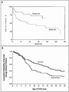
Fig. 2. Survival in breastfed versus bottle-fed HIV-infected infants
(A) Survival to 36 months among HIV-infected infants from diagnosis by type of feeding, 64 bottle fed infants (dotted line) versus 36 breast-fed infants (solid line) from the Italian National Registry of AIDS through February 1990 (Breslow p=0.01; Mantel-Cox p=0.003) from Tozzi et al. (240). (B) Survival to 24 months among children who had HIV infection by 4 months of age (71 children assigned to abrupt weaning at 4 months and 81 assigned to the control group of standard breastfeeding practices with median duration of breastfeeding of 16 months) from a controlled trial in Zambia (p = 0.007) from Kuhn et al (40).
Fig. 3
Fig. 3. Relative levels of HIV RNA (red) and CD4 cells (blue) in adults (dotted lines) and children (solid lines) in the years following acquisition of HIV-1
Gray boxes highlight the median time to AIDS/Death for infants by region and route of infection in comparison to adults. US – United States.
Fig. 4
Fig. 4. Distribution of follow-up and events within age groups, for analyses of (A) CD4% and death, (B) CD4% and AIDS, (C) viral load and death, (D) viral load and AIDS by age from the HIV Paediatric Prognostic Markers Collaborative Study Group (101)
Fig. 5
Fig. 5. Death rate per 100 person-years by current CD4+ T-cell count and age
Children aged 0–4 years of age have markedly decreased survival even at high CD4 cell counts whereas adolescent and young adults (15–24 years) have a survival advantage. Adapted from Dunn et al (107).
Fig. 6
Fig. 6. T-cell populations (Panel A) and CD4+ T-cell population subsets (Panel B) by age
(A) CD4+ T lymphocytes in blue, CD8+ T lymphocytes in black, and CD19+ B lymphocytes in red by number of cells (solid lines) and percentage of lymphocytes (dotted lines) by age. (B) Tcm = central memory T cells (CD3+CD4+CD45RACD27+) in red, CXCR5 = CXCR5+ memory helper T cells (CD3+CD4+CD45RO+CD185+) in blue, and Tregs = regulatory T cells (CD3+CD4+CD25+CD127) in black by number of cells (solid lines) and percentage of CD4+ T cells (dotted lines) by age. Adapted from Schartorje et al. (136).
Fig. 7
Fig. 7. Percent of adult immunoglobulin levels by age for IgG (blue), IgM (green), and IgA (red) showing significant reduction in immunoglobulin levels in children persisting into adolescence
Adapted from Stiehm (138).
Fig. 8
Fig. 8. Development of gut and its immune system with appearance of cellular factors by gestational age
From Insoft et al. (147).

References

    1. Rates of mother-to-child transmission of HIV-1 in Africa, America, and Europe: results from 13 perinatal studies. The Working Group on Mother-To-Child Transmission of HIV. J Acquir Immune Defic Syndr Hum Retrovirol. 1995;8:506–510. - PubMed
    1. Taha TE, et al. Association of recent HIV infection and in-utero HIV-1 transmission. AIDS. 2011;25:1357–1364. - PMC - PubMed
    1. Aldrovandi GM, Kuhn L. What infants and breasts can teach us about natural protection from HIV infection. J Infect Dis. 2010;202(Suppl):S366–S370. - PMC - PubMed
    1. Adams KM, Nelson JL. Microchimerism: an investigative frontier in autoimmunity and transplantation. JAMA. 2004;291:1127–1131. - PubMed
    1. Lee TH, Chafets DM, Biggar RJ, McCune JM, Busch MP. The role of transplacental microtransfusions of maternal lymphocytes in in utero HIV transmission. J Acquir Immune Defic Syndr. 2010;55:143–147. - PMC - PubMed

Publication types