Review
. 2013 Jun;3(3):296-306.
doi: 10.1016/j.coviro.2013.05.019. Epub 2013 Jun 15.

Viral modulation of programmed necrosis

Affiliations
Review

Viral modulation of programmed necrosis

William J Kaiser et al. Curr Opin Virol. 2013 Jun.

Abstract

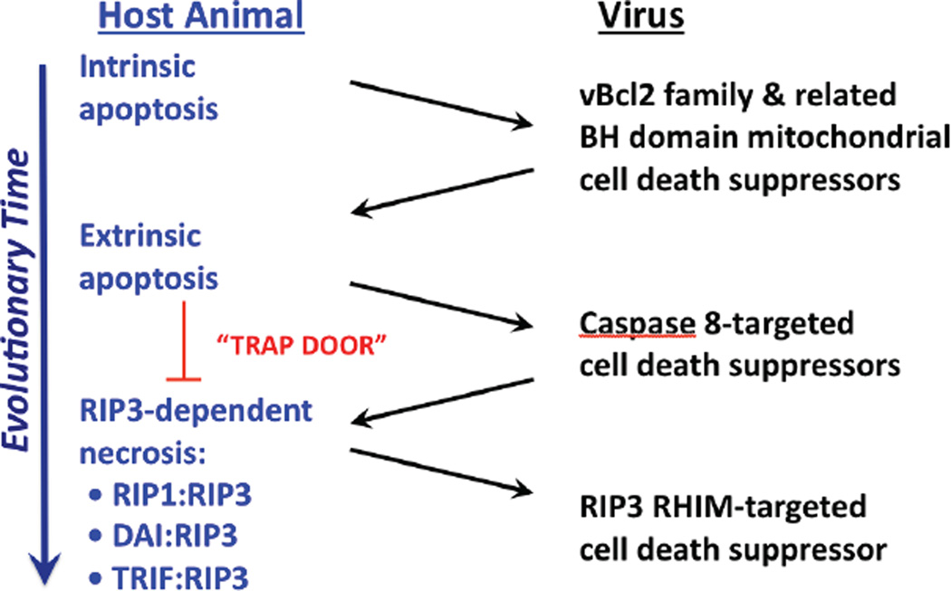
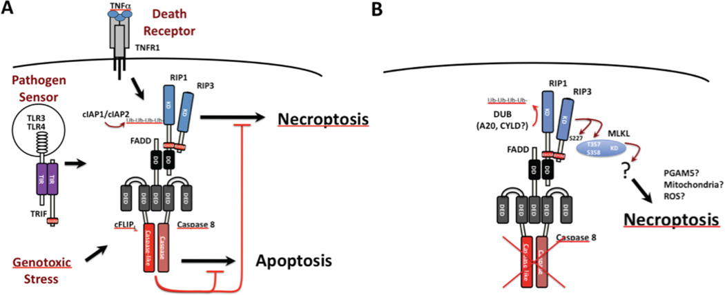
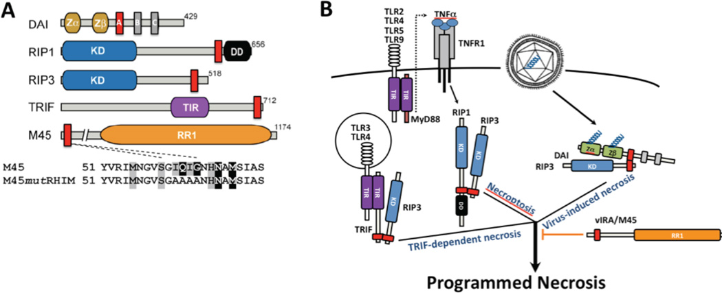
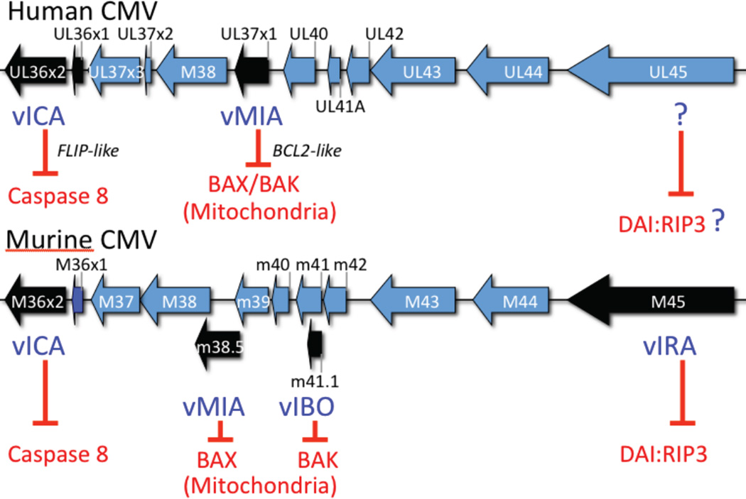
Apoptosis and programmed necrosis balance each other as alternate first line host defense pathways against which viruses have evolved countermeasures. Intrinsic apoptosis, the critical programmed cell death pathway that removes excess cells during embryonic development and tissue homeostasis, follows a caspase cascade triggered at mitochondria and modulated by virus-encoded anti-apoptotic B cell leukemia (BCL)2-like suppressors. Extrinsic apoptosis controlled by caspase 8 arose during evolution to trigger executioner caspases directly, circumventing viral suppressors of intrinsic (mitochondrial) apoptosis and providing the selective pressure for viruses to acquire caspase 8 suppressors. Programmed necrosis likely evolved most recently as a 'trap door' adaptation to extrinsic apoptosis. Receptor interacting protein (RIP)3 kinase (also called RIPK3) becomes active when either caspase 8 activity or polyubiquitylation of RIP1 is compromised. This evolutionary dialog implicates caspase 8 as a 'supersensor' alternatively activating and suppressing cell death pathways.


Figures

Figure 1
Figure 1. Evolution of cell death pathways in host defense
Diagram representing evolutionary time (large arrow) together with host death pathways targeted by known virus-encoded cell death suppressors. Intrinsic apoptosis is a primordial property of multicellular organisms to eliminate excess cells during development that contributes to host defense. Herpesviruses, poxviruses and adenoviruses encode mitochondrial cell death suppressors that limit the impact of pro-apoptotic BCL2 family member signaling in host defense [–6,13]. Extrinsic apoptosis represents an adaptation to avoid virus-encoded inhibitors of mitochondrial apoptosis, by employing Casp8 as a self-activating protease that can directly target executioner caspases, Casp3 and Casp7. Herpesviruses, poxviruses and adenoviruses acquired suppressors of Casp8 that limit the contribution of extrinsic apoptosis to elimination of virus-infected cells. Programmed necrosis represents an adaptation to take advantage of Casp8 inhibition, by opening a trap door via any of three signaling platforms, RIP1-RIP3, DAIRIP3 and TRIF-RIP3. Some viruses, such as vaccinia, remain susceptible to programmed necrosis; whereas, other viruses, such as murine CMV, encode suppressors that limit the impact of this pathway.
Figure 2
Figure 2. Caspase 8 regulation of extrinsic apoptosis and programmed necrosis
A. Diagrammatic summary of the cytoprotective FADD-Casp8-cFLIPL-RIP1-RIP3 signaling complex (complex IIb or ripoptosome). The cytosolic complex forms downstream of cell surface death receptor (e.g. TNFR1 signaling) [87,117,118], endosomal pathogen sensor (e.g. TLR3/TLR4 signaling) [60,152] or intracellular genotoxic stress [61]. The cFLIPL-Casp8 heterodimer association with FADD prevents self-cleavage activation that is necessary to initiate apoptosis while maintaining sufficient basal activity to prevent unleashed RIP1-RIP3 necroptosis. E3 ubiquitin (Ub) ligases cIAP1 and cIAP2 also prevent RIP1-RIP3 necroptosis by adding K63 Ub chains to RIP1 and possibly other targets [61,62,69]. B. Diagrammatic summary of necroptosis unleashed by RIP3. In the presence of caspase compromise (red “X”) or when cIAP1 and cIAP2 are compromised and de-ubiquitinases (DUBs) such as A20 and CYLD predominate and eliminate poly Ub chains, RIP3 kinase becomes activated and is essential for activating MLKL kinase activity [76], autophosphorylating at S277 and also phosphorylating T357 and S358 on MLKL [78] to promote the direct interaction between these two kinases, an essential step in necroptosis.
Figure 3
Figure 3. Signaling adaptors in control of RIP3 programmed necrosis
A. Diagrammatic depiction of host and viral RHIM-containing adaptors. Mouse DAI [148,150], RIP1 [153], RIP3 [154,155] and TRIF [145,146], together with murine CMV M45-encoded vIRA, an RR1 homolog [126]. The expanded regions depicts RHIM region from aa 51 and the tetra-alanine mutation employed to characterize M45 RHIM-specific inhibition [89,140]. Kinase domain (KD) of RIP1 and RIP3, death domain (DD) of RIP1, toll-IL-1 receptor (TIR) domain of TRIF and RIP homotypic interaction motif (RHIM; red boxes). DAI has three RHIM-like repeat (RLR) elements where RLR A is functional [148]. B. Summary diagram depicting three distinct triggers of RIP3 kinase-dependent programmed necrosis. TNFR1-induced RIP1-RIP3 necroptosis [87,117,118], DAI-RIP3 virus-induced necrosis [89,93] and TLR3 or TRL4 TRIF-dependent necrosis, as well as TLR2, TLR4, TLR5 and TLR9 MyD88-dependent activation of TNF and secondary induction of necroptosis [94]. These complexes depend on RHIM interactions and are blocked by murine CMV M45-encoded vIRA [89,93,94].
Figure 4
Figure 4. Cytomegalovirus cell death suppression locus
Colinear regions of the human and murine CMV genomes showing the location of ORFs that encode cell death suppressors. vICA is a sequence homolog lacking a DED that nevertheless functions in a FLIP-like manner, interacting with Casp8 to prevent self-activation [30,31]. Human CMV encoded vMIA inhibits BAX [: Poncet, 2004 #5807] and modulates BAK [157], whereas murine CMV encodes vMIA to inhibit BAX and vIBO to inhibit BAK at mitochondria. Murine CMV M45-encoded vIRA suppresses apoptosis and necrosis [,–140], acting as a RHIM inhibitor to suppress DAIRIP3 complex formation [93] (see Fig. 3).

References

    1. Danial NN, Korsmeyer SJ. Cell death: critical control points. Cell. 2004;116:205–219. The classic review of intrinsic cell death reliance on BCL2 family members.

    1. Kroemer G, Galluzzi L, Brenner C. Mitochondrial membrane permeabilization in cell death. Physiol Rev. 2007;87:99–163. - PubMed
    1. Tait SW, Green DR. Mitochondria and cell signalling. J Cell Sci. 2012;125:807–815. - PMC - PubMed
    1. Everett H, McFadden G. Apoptosis: an innate immune response to virus infection. Trends Microbiol. 1999;7:160–165. - PubMed
    1. Benedict CA, Norris PS, Ware CF. To kill or be killed: viral evasion of apoptosis. Nat Immunol. 2002;3:1013–1018. - PubMed

Publication types