

Multiple peripheral typical carcinoid tumors of the lung: associated with sclerosing hemangiomas
- PMID: 23773456
- PMCID: PMC3728223
- DOI: 10.1186/1746-1596-8-97
Multiple peripheral typical carcinoid tumors of the lung: associated with sclerosing hemangiomas
Abstract
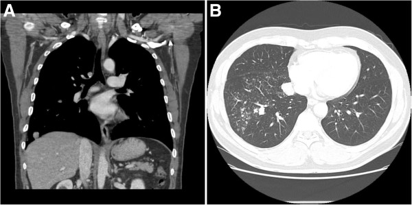
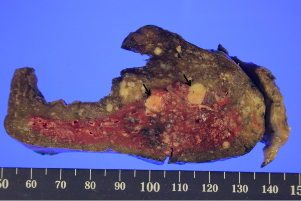
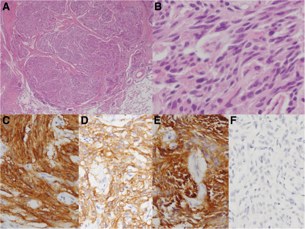
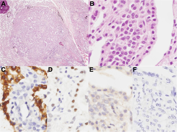
This study presents a first case of multiple peripheral typical carcinoid tumors associated with sclerosing hemangiomas in the lung. A 52-year-old male presented with incidentally detected multiple pulmonary nodules on a simple chest X-ray during routine health check-up. A computed tomography (CT) scan of the chest showed multiple nodular lesions in the middle and lower lobes of the right lung. These were initially suspected as inflammatory lesions due to miliary tuberculosis. However, possibility of malignancy could not be excluded and right lower lobe lobectomy was performed. Histopathologically, some nodules including two largest nodules were composed of small round to spindle shaped cells with fine chromatin pattern, whereas the rest of the sclerotic nodules were composed of two epithelial cell types- surface cells and round cells. The final diagnosis of this case was multiple peripheral typical carcinoid tumors associated with sclerosing hemangiomas of the lung. For past three years of post-surgery follow up period, no new lesions or changes in the right middle lobe have been identified.
Figures
Similar articles
-
Sclerosing pneumocytoma mixed with a typical carcinoid tumor: A case report and review of literature.Medicine (Baltimore). 2019 Feb;98(5):e14315. doi: 10.1097/MD.0000000000014315. Medicine (Baltimore). 2019. PMID: 30702609 Free PMC article. Review.
-
Bilateral multiple sclerosing hemangiomas of the lung.Gen Thorac Cardiovasc Surg. 2009 Dec;57(12):667-70. doi: 10.1007/s11748-009-0452-y. Gen Thorac Cardiovasc Surg. 2009. PMID: 20013104
-
Coexistence of pulmonary sclerosing hemangioma and primary adenocarcinoma in the same nodule of lung.Diagn Pathol. 2011 May 20;6:41. doi: 10.1186/1746-1596-6-41. Diagn Pathol. 2011. PMID: 21599956 Free PMC article.
-
CT features of peripheral pulmonary carcinoid tumors.AJR Am J Roentgenol. 2011 Nov;197(5):1073-80. doi: 10.2214/AJR.10.5954. AJR Am J Roentgenol. 2011. PMID: 22021498
-
Sclerosing hemangioma of the lung: a clinicopathologic study.Zhonghua Yi Xue Za Zhi (Taipei). 1993 Sep;52(3):149-54. Zhonghua Yi Xue Za Zhi (Taipei). 1993. PMID: 8252456 Review.
Cited by
-
Sclerosing pneumocytoma mixed with a columnar clear cell adenoma and a typical carcinoid: case report and review of literature.Int J Clin Exp Pathol. 2020 Oct 1;13(10):2599-2607. eCollection 2020. Int J Clin Exp Pathol. 2020. PMID: 33165428 Free PMC article.
-
Sclerosing pneumocytoma mixed with a typical carcinoid tumor: A case report and review of literature.Medicine (Baltimore). 2019 Feb;98(5):e14315. doi: 10.1097/MD.0000000000014315. Medicine (Baltimore). 2019. PMID: 30702609 Free PMC article. Review.
-
Automated quantification of Ki-67 proliferative index of excised neuroendocrine tumors of the lung.Diagn Pathol. 2014 Oct 16;9:174. doi: 10.1186/s13000-014-0174-z. Diagn Pathol. 2014. PMID: 25318848 Free PMC article.
-
Multidisciplinary management of advanced lung neuroendocrine tumors.J Thorac Dis. 2015 Apr;7(Suppl 2):S163-71. doi: 10.3978/j.issn.2072-1439.2015.04.20. J Thorac Dis. 2015. PMID: 25984363 Free PMC article. Review.
-
Multifocal neuroendocrine cell hyperplasia accompanied by tumorlet formation and pulmonary sclerosing pneumocytoma: A case report.World J Clin Cases. 2020 Aug 26;8(16):3583-3590. doi: 10.12998/wjcc.v8.i16.3583. World J Clin Cases. 2020. PMID: 32913868 Free PMC article.
References
-
- Ttravis WD, Brambilla W, Muller-Hermelink HK, Harris CC. World health organization classification of tumors: pathology and genetics of tumors of the lung, pleura, thymus and heart. ICRC Press; 2004.
-
- Nicholson AG, Magkou C, Snead D, Vohra HA, Sheppard MN, Goldstraw P, Beddow E, Hansell DM, Travis WD, Corrin B. Unusual sclerosing haemangiomas and sclerosing haemangioma-like lesions, and the value of TTF-1 in making the diagnosis. Histopathology. 2002;41:404–413. doi: 10.1046/j.1365-2559.2002.01522.x. - DOI - PubMed
-
- Suzuki K, Shiono S, Kato H, Yanagawa N, Sato T. [Small sclerosing hemangioma combined with primary lung cancer; report of a case] Kyobu Geka. 2006;59:590–593. - PubMed
-
- Schiergens TS, Khalil PN, Mayr D, Thasler WE, Angele MK, Hatz RA, Jauch KW, Kleespies A. Pulmonary sclerosing hemangioma in a 21-year-old male with metastatic hereditary non-polyposis colorectal cancer: report of a case. World J Surg Oncol. 2011;9:62. doi: 10.1186/1477-7819-9-62. - DOI - PMC - PubMed
Publication types
MeSH terms
LinkOut - more resources
Full Text Sources
Other Literature Sources
Medical
Miscellaneous
