. 2014 Jan;20(1):64-71.
doi: 10.1177/1352458513492244. Epub 2013 Jun 17.

Perivenular brain lesions in a primate multiple sclerosis model at 7-tesla magnetic resonance imaging

Affiliations

Perivenular brain lesions in a primate multiple sclerosis model at 7-tesla magnetic resonance imaging

María I Gaitán et al. Mult Scler. 2014 Jan.

Abstract

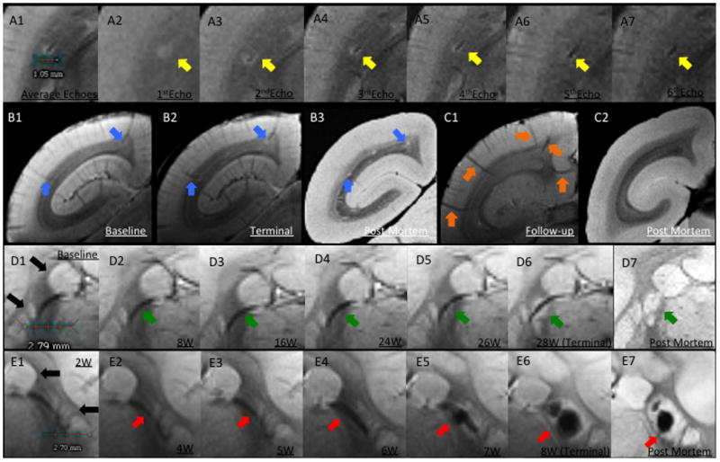
BACKGROUND Magnetic resonance imaging (MRI) can provide in vivo assessment of tissue damage, allowing evaluation of multiple sclerosis (MS) lesion evolution over time--a perspective not obtainable with postmortem histopathology. Relapsing-remitting experimental autoimmune encephalomyelitis (EAE) is an experimental model of MS that can be induced in the common marmoset, a small new world primate, and that causes perivenular white matter (WM) lesions similar to those observed in MS. METHODS Brain lesion development and evolution were studied in vivo and postmortem in four marmosets with EAE through serial T2- and T2*-weighted scans at 7-tesla. Supratentorial WM lesions were identified and characterized. RESULTS Of 97 lesions observed, 86 (88%) were clearly perivenular, and 62 (72%) developed around veins that were visible even prior to EAE induction. The perivenular configuration was confirmed by postmortem histopathology. Most affected veins, and their related perivascular Virchow-Robin spaces, passed into the subarachnoid space rather than the ventricles. CONCLUSION As in human MS, the intimate association between small veins and EAE lesions in the marmoset can be studied with serial in vivo MRI. This further strengthens the usefulness of this model for understanding the process of perivenular lesion development and accompanying tissue destruction in MS.

Keywords: Veins; experimental autoimmune encephalomyelitis; magnetic resonance imaging; multiple sclerosis; susceptibility weighted imaging.


Conflict of interest statement

Disclosure/Conflict of Interest: The authors have no conflicts of interest to disclose.

Figures

Figure 1
Figure 1
Lesion and vein characteristics. A. Cumulative supratentorial lesion volume vs. time for all marmosets, two of which were immunized with recombinant human myelin oligodendrocyte glycoprotein (rhMOG) and two with human white matter homogenate (WMH). B. Supratentorial WM Total number of lesions, number of perivenular lesions, and number of perivenular lesions for which pre-existing veins could be detected on the baseline MRI. C. Vein topography. Number and location (deep vs. superficial) of veins involved in lesions. D. Number of lesions with deep cerebral veins (total and pre-existing). E. Number of lesions with superficial cerebral veins (total and pre-existing).
Figure 2
Figure 2
Perivenular white matter lesion. A. Ex vivo T2* MRI. Note that after brain perfusion the central vein is not well seen; this is due to replacement of blood with perfusate, which is invisible on MRI. B. Luxol-fast blue staining for myelin. The demyelinated lesion is centered on a small vein (yellow arrow), which is surrounded by a thick inflammatory cuff (orange arrow). C. In vivo averaged T2* showing the hypointense central vein. D. Ex vivo T2* MRI. Note that after brain perfusion the blood is not replaced with perfusate. E. Hematoxilin and Eosin staining. Acute area of hemorrhage – note the extravascular red blood cells. F. Perls' Prussian blue stainin. The staining is negative for iron deposition.
Figure 3
Figure 3
Perivenular development of lesions. A. In vivo T2*. A1, Average of all 6 echoes: A2, TE (echo time)=3.5ms; A3, TE=10.5ms; A4, TE=17.5ms; A5, TE=24.5ms; A6, TE=31.5ms; A7, TE=38.5ms. In A1, the dark line represents the lesion's central vessel, which is surrounded by the bright lesion. A2-A7, Progressive darkening of the central vessel relative to the surrounding lesion, reflecting increasing magnetic susceptibility. B. Left brain hemisphere of WMH1. B1, Arrows point to two pre-existing superficial veins visible at baseline, before EAE induction; B2, Lesions surround these two veins on the final scan; B3, Removal of intravascular blood at time of perfusion allows clear visualization of lesions, but not associated veins, on postmortem MRI. C, Left hemisphere of a control animal. C1, Arrows point to five representative superficial veins; C2, Postmortem MRI with nonvisualized superficial veins. D. Lesion development around a deep white matter vein in WMH2. D1, Baseline (pre-immunization) T2* average. Note that basal ganglia structures have similar signal intensity to lesions (black arrows). D2-D6, Consecutive averaged T2*-weighted images at different time points (W=weeks after immunization). Arrows point to a deep vein that was detected at baseline and was unchanged until 24 weeks, when the earliest sign of the developing lesion could be detected. D7, Postmortem T2*, showing the apparent absence of a central vein due to removal of intravascular blood. E. Lesion development surrounding a deep white matter vein in rhMOG1, with eventual hemorrhage. E1-E6, Consecutive averaged T2*-weighted images at different time points. E5-E6, Arrows point to a lesion with central hemorrhage. E7, Postmortem MRI shows persistent central low signal related to the intralesional hemorrhage. Similar to D, the vein itself is not visualized on the postmortem scan due to removal of intravascular blood.

References

    1. Lucchinetti C, Bruck W, Parisi J, Scheithauer B, Rodriguez M, Lassmann H. Heterogeneity of multiple sclerosis lesions: implications for the pathogenesis of demyelination. Annals of neurology. 2000;47(6):707–17. Epub 2000/06/14. - PubMed
    1. Charcot JM. Histologie de la sclérose en plaques. Paris: Imprimerie L. Pupart-Davyl; 1869. p. 23.
    1. Tallantyre EC, Brookes MJ, Dixon JE, Morgan PS, Evangelou N, Morris PG. Demonstrating the perivascular distribution of MS lesions in vivo with 7-Tesla MRI. Neurology. 2008;70(22):2076–8. Epub 2008/05/29. - PubMed
    1. Adams CW. The onset and progression of the lesion in multiple sclerosis. Journal of the neurological sciences. 1975;25(2):165–82. Epub 1975/06/01. - PubMed
    1. Henderson AP, Barnett MH, Parratt JD, Prineas JW. Multiple sclerosis: distribution of inflammatory cells in newly forming lesions. Ann Neurol. 2009;66(6):739–53. Epub 2009/12/26. - PubMed

Publication types