. 2013 Sep;81(9):3182-97.
doi: 10.1128/IAI.00291-13. Epub 2013 Jun 17.

Production and release of antimicrobial and immune defense proteins by mammary epithelial cells following Streptococcus uberis infection of sheep

Affiliations

Production and release of antimicrobial and immune defense proteins by mammary epithelial cells following Streptococcus uberis infection of sheep

Maria Filippa Addis et al. Infect Immun. 2013 Sep.

Abstract

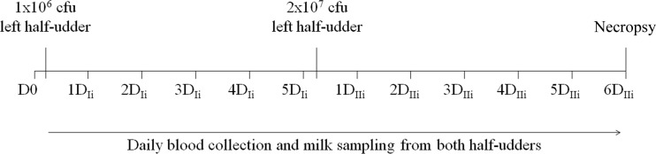
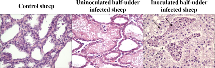
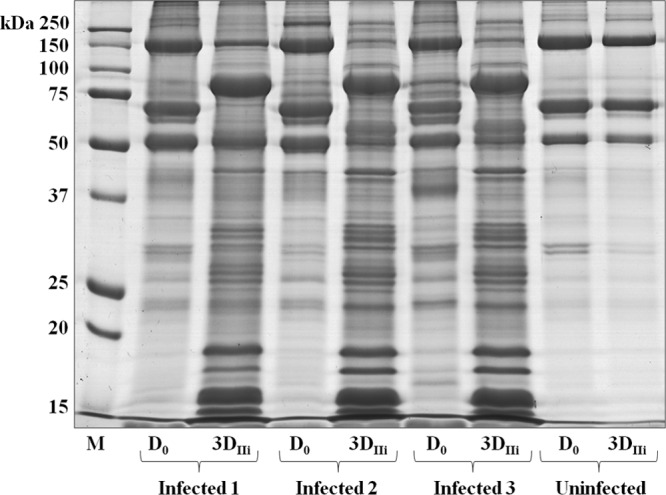
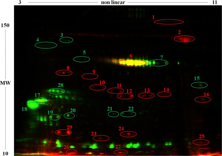
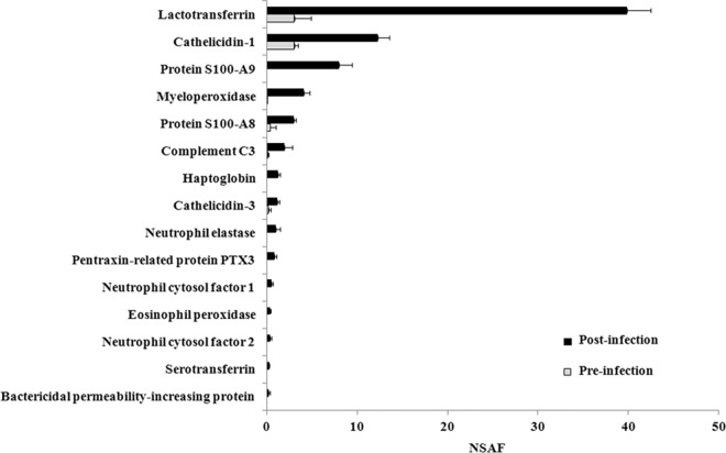
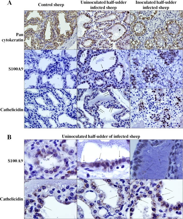
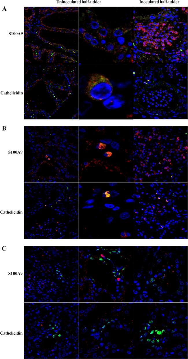
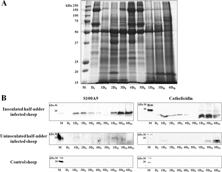
Investigating the innate immune response mediators released in milk has manifold implications, spanning from elucidation of the role played by mammary epithelial cells (MECs) in fighting microbial infections to the discovery of novel diagnostic markers for monitoring udder health in dairy animals. Here, we investigated the mammary gland response following a two-step experimental infection of lactating sheep with the mastitis-associated bacterium Streptococcus uberis. The establishment of infection was confirmed both clinically and by molecular methods, including PCR and fluorescent in situ hybridization of mammary tissues. Proteomic investigation of the milk fat globule (MFG), a complex vesicle released by lactating MECs, enabled detection of enrichment of several proteins involved in inflammation, chemotaxis of immune cells, and antimicrobial defense, including cathelicidins and calprotectin (S100A8/S100A9), in infected animals, suggesting the consistent involvement of MECs in the innate immune response to pathogens. The ability of MECs to produce and release antimicrobial and immune defense proteins was then demonstrated by immunohistochemistry and confocal immunomicroscopy of cathelicidin and the calprotectin subunit S100A9 on mammary tissues. The time course of their release in milk was also assessed by Western immunoblotting along the course of the experimental infection, revealing the rapid increase of these proteins in the MFG fraction in response to the presence of bacteria. Our results support an active role of MECs in the innate immune response of the mammary gland and provide new potential for the development of novel and more sensitive tools for monitoring mastitis in dairy animals.


Figures

Fig 1
Fig 1
Timeline of experimental infection: sample collection, S. uberis inoculation, and animal sacrifice and necropsy.
Fig 2
Fig 2
Histopathological grading. Representative hematoxylin-eosin-stained sections of tissue from control and experimentally infected sheep are shown. The tissues of the control sheep and uninoculated half udders of infected sheep are histologically normal. Inoculated half udders show intra-alveolar neutrophils (solid arrow) and signs of degeneration of the secretory epithelium (dashed arrows). Magnifications, ×200.
Fig 3
Fig 3
SDS-PAGE of sheep MFGPs before and after infection with S. uberis. The image illustrates a representative gel of all proteins extracted from MFGs isolated from the milk of all animals before infection (D0) and 3 days after the second inoculation (3DIIi). Lane M, molecular mass markers.
Fig 4
Fig 4
Representative 2D DIGE profile of MFGPs from sheep before (green) and after (red) infection with S. uberis. An overlay image of representative MFGP profiles obtained before (green) and after (red) infection with S. uberis is shown. Yellow spots result from the superimposition of red and green signals and indicate similar levels of protein expression. Spots showing statistically significant differences are circled, and identities according to MS analysis are reported in Table 2. MW, molecular weight (molecular weights are given in thousands).
Fig 5
Fig 5
Normalized spectral abundance factors (NSAFs) of selected antimicrobial proteins. Bars indicate the abundance and standard deviation abundance of antimicrobial proteins in the milk fat fraction before and after infection. Proteins having direct antimicrobial functions according to the Uniprot Knowledgebase and showing statistically significant changes in abundance (RSC) between the two conditions (Table 3) are reported.
Fig 6
Fig 6
Immunohistochemical analysis of mammary tissues. (A) Reactivity of mammary tissues from the indicated animals for pancytokeratin, S100A9, and cathelicidin. (B) Reactivity of mammary tissues from the uninoculated half udders of infected animals for S100A9 and cathelicidin. All animals included in the experiment were tested in replicate experiments. Arrows, focal points of reactivity.
Fig 7
Fig 7
Immune colocalization analysis of mammary tissues. The reactivities of mammary tissues to antibodies against S100A9 and cathelicidin (green signals), together with cell-type-specific antibodies (red signals), are shown. (A) Epithelial cells; (B) neutrophils; (C) macrophages.
Fig 8
Fig 8
Results of FISH for S. uberis. The reactivity obtained with an S. uberis-specific probe is shown in the mammary tissues of the control sheep, the uninoculated half udders of infected animals, and the inoculated half udders of infected animals.
Fig 9
Fig 9
Time course of the MFGP profile and of S100A9 and cathelicidin reactivity over the course of infection. (A). Total MFGP profile over the course of infection, starting with preinoculation (D0) and continuing to the last day of the second inoculation with S. uberis (6DIIi). (B) Reactivity of S100A9 and cathelicidin in inoculated and uninoculated half udders of infected animals and in control animals. Lanes M, molecular mass markers.

Similar articles

Cited by

References

    1. Zadoks RN, Middleton JR, McDougall S, Katholm J, Schukken YH. 2011. Molecular epidemiology of mastitis pathogens of dairy cattle and comparative relevance to humans. J. Mammary Gland Biol. Neoplasia 16:357–372 - PMC - PubMed
    1. De Vliegher S, Fox LK, Piepers S, McDougall S, Barkema HW. 2012. Mastitis in dairy heifers: nature of the disease, potential impact, prevention, and control. J. Dairy Sci. 95:1025–1040 - PubMed
    1. Marogna G, Rolesu S, Lollai S, Tola S, Leori S. 2010. Clinical findings in sheep farms affected by recurrent bacterial mastitis. Small Rumin. Res. 88:119–125
    1. Rainard P, Riollet C. 2006. Innate immunity of the bovine mammary gland. Vet. Res. 37:369–400 - PubMed
    1. Beutler B. 2004. Innate immunity: an overview. Mol. Immunol. 40:845–859 - PubMed

Publication types

MeSH terms