Review
. 2013 Aug;9(8):459-69.
doi: 10.1038/nrneph.2013.110. Epub 2013 Jun 18.

Contribution of aldosterone to cardiovascular and renal inflammation and fibrosis

Affiliations
Review

Contribution of aldosterone to cardiovascular and renal inflammation and fibrosis

Nancy J Brown. Nat Rev Nephrol. 2013 Aug.

Abstract

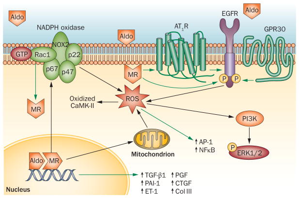
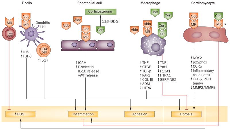
The steroid hormone aldosterone regulates sodium and potassium homeostasis. Aldosterone and activation of the mineralocorticoid receptor also causes inflammation and fibrosis of the heart, fibrosis and remodelling of blood vessels and tubulointerstitial fibrosis and glomerular injury in the kidney. Aldosterone and mineralocorticoid-receptor activation initiate an inflammatory response by increasing the generation of reactive oxygen species by nicotinamide adenine dinucleotide phosphate (NADPH) oxidase and mitochondria. High salt intake potentiates these effects, in part by activating the Rho family member Rac1, a regulatory subunit of reduced NADPH oxidase that activates the mineralocorticoid receptor. Studies in mice in which the mineralocorticoid receptor has been deleted from specific cell types suggest a key role for macrophages in promoting inflammation and fibrosis. Aldosterone can exert mineralocorticoid-receptor-independent effects via the angiotensin II receptor and via G-protein-coupled receptor 30. Mineralocorticoid-receptor antagonists are associated with decreased mortality in patients with heart disease and show promise in patients with kidney injury, but can elevate serum potassium concentration. Studies in rodents genetically deficient in aldosterone synthase or treated with a pharmacological aldosterone-synthase inhibitor are providing insight into the relative contribution of aldosterone compared with the contribution of mineralocorticoid-receptor activation in inflammation, fibrosis, and injury. Aldosterone-synthase inhibitors are under development in humans.


Conflict of interest statement

Competing interests

The author declares associations with the following companies: Novartis, Shire Pharmaceuticals. See the article online for full details of the relationships.

Figures

Figure 1
Figure 1
Mechanisms by which aldosterone and/or MR activation induce oxidative stress, inflammation, and fibrosis. Activation of the MR by aldosterone results in dissociation of chaperone proteins, translocation of the MR to the nucleus and activation of transcription. MR activation induces oxidative stress by both NADPH oxidase and mitochondria. ROS formed by NADPH oxidase oxidize CaMK-II. Oxidative stress triggers the activation of proinflammatory transcription factors such as AP-1 and NFκB. Rac1 can also activate the mineralocorticoid receptor. Aldosterone can induce rapid, nongenomic effects via GPR30. Aldosterone and angiotensin II interact to cause rapid, nongenomic effects via transactivation of the EGFR, resulting in the generation of ROS and phosphorylation of ERK1/2. The green arrows indicate an activating or increasing effect; the black arrows indicate a downstream consequence. See text for references. Abbreviations: Aldo, aldosterone; AP-1, activator protein 1; AT1R, angiotensin II type 1 receptor; CAMK-II, calcium/calmodulin-dependent protein kinase type II subunit gamma; Coll III, collagen III; CTGF, connective tissue growth factor; EGFR, epidermal growth factor receptor; ERK1/2, extracellular signal-regulated kinases 1 and 2 (ERK)1/2; ET-1, endothelin 1; GPR30, G-protein-coupled receptor 30; MR, mineralocorticoid receptor; NADPH, nicotinamide adenine dinucleotide phosphate; NFκB, nuclear factor kappa B; P, phosphate; PAI-1, plasminogen activator inhibitor 1; PGF, placental growth factor; PI3K, phosphatidylinositol 3-kinase; ROS, reactive oxygen species; TGF-β1, transforming growth factor-β1.
Figure 2
Figure 2
Effects of cell-specific activation of the MR. Aldosterone activates the MR on macrophages to produce inflammation, hypertrophy, and fibrosis in the heart. At low concentrations, cortisol can also cause an inflammatory response via activation of glucocorticoid receptors. At higher concentrations, cortisol induces alternative activation of macrophages. Activation of MRs in cardiomyocytes increases the formation of ROS and might promote fibrosis. Studies in which 11βHSD-2 is overexpressed in cardiomyocytes suggest that cortisol opposes these effects. In endothelial cells, MR activation by aldosterone promotes inflammation and adhesion of leukocytes; endothelial cells produce 11βHSD-2, rendering cortisol inactive at the mineralocorticoid receptor. Stimulation of the MR on dendritic cells increases production of IL-6 and TGF-β1, primes CD8+ T cells, and induces TH17 production by CD4+ cells. The dotted line indicates that the positive effect is not consistent across studies (see text). The red lines indicate a decreasing effect. See text for references. Abbreviations: 11βHSD-2, 11β-hydroxysteroid dehydrogenase type II; ADM, adrenomedullin; Aldo, aldosterone; CCR5, C-C chemokine receptor type 5; Cort, cortisol; COL III, collagen III; CTGF, connective tissue growth factor; F13A1, coagulation factor XIII, A1 polypeptide; GR, glucocorticoid receptor; HTRA, high temperature requirement factor A1; ICAM1, intercellular adhesion molecule 1; IL, interleukin; MMP, matrix metalloproteinase; MR, mineralocorticoid receptor; PAI-1, plasminogen activator inhibitor 1; ROS, reactive oxygen species; TGF-β, transforming growth factor-β; TNF, tumour necrosis factor; TREG, regulatory T cell; vWF, von Willebrand factor; Ym1, a secretory protein produced by activated macrophages.

References

    1. Náray-Fejes-Tóth A, Canessa C, Cleaveland ES, Aldrich G, Fejes-Tóth G. sgk is an aldosterone-induced kinase in the renal collecting duct Effects on epithelial na+ channels. J Biol Chem. 1999;274:16973–16978. - PubMed
    1. Snyder PM, Olson DR, Thomas BC. Serum and glucocorticoid-regulated kinase modulates Nedd4-2-mediated inhibition of the epithelial Na+ channel. J Biol Chem. 2002;277:5–8. - PubMed
    1. Bhalla V, Soundararajan R, Pao AC, Li H, Pearce D. Disinhibitory pathways for control of sodium transport: regulation of ENaC by SGK1 and GILZ. Am J Physiol Renal Physiol. 2006;291:F714–F721. - PubMed
    1. Rozansky DJ, et al. Aldosterone mediates activation of the thiazide-sensitive Na-Cl cotransporter through an SGK1 and WNK4 signaling pathway. J Clin Invest. 2009;119:2601–2612. - PMC - PubMed
    1. Arroyo JP, et al. Nedd4–2 modulates renal Na+-Cl− cotransporter via the aldosterone-SGK1-Nedd4–2 pathway. J Am Soc Nephrol. 2011;22:1707–1719. - PMC - PubMed

Publication types

MeSH terms