. 2013 Jul 26;288(30):21667-77.
doi: 10.1074/jbc.M113.480657. Epub 2013 Jun 17.

Ca2+-dependent structural changes in the B-cell receptor CD23 increase its affinity for human immunoglobulin E

Affiliations

Ca2+-dependent structural changes in the B-cell receptor CD23 increase its affinity for human immunoglobulin E

Daopeng Yuan et al. J Biol Chem. .

Abstract

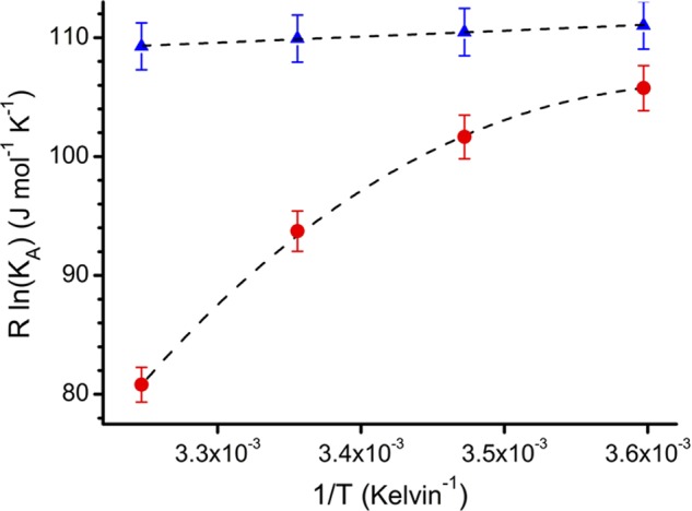
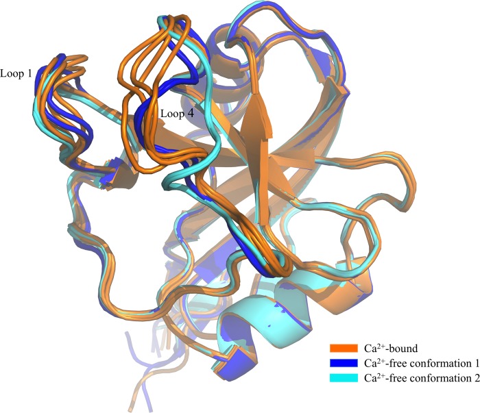
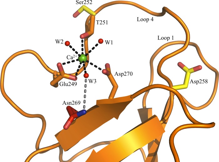
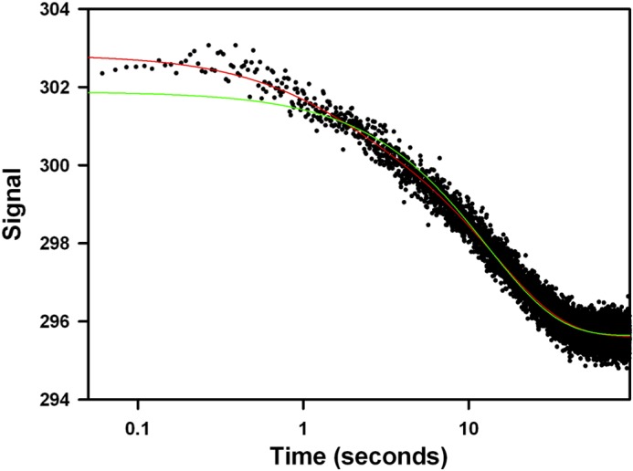
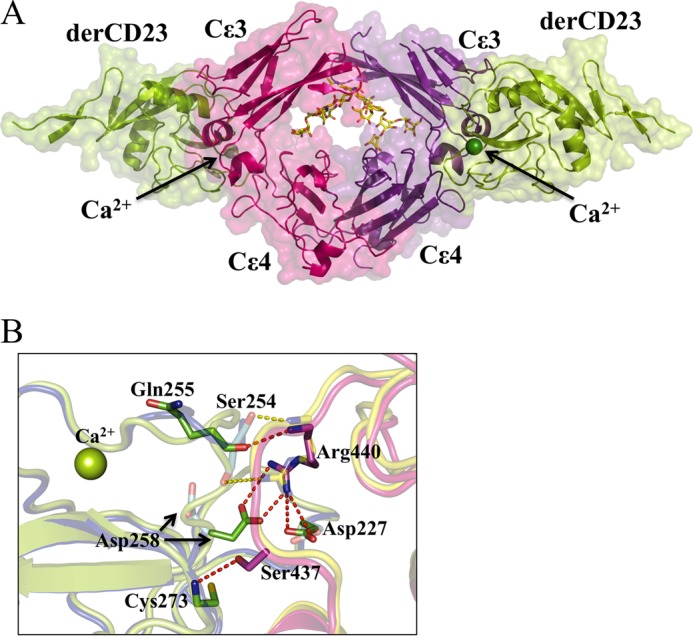
Immunoglobulin E (IgE) antibodies play a fundamental role in allergic disease and are a target for therapeutic intervention. IgE functions principally through two receptors, FcεRI and CD23 (FcεRII). Minute amounts of allergen trigger mast cell or basophil degranulation by cross-linking IgE-bound FcεRI, leading to an inflammatory response. The interaction between IgE and CD23 on B-cells regulates IgE synthesis. CD23 is unique among Ig receptors in that it belongs to the C-type (calcium-dependent) lectin-like superfamily. Although the interaction of CD23 with IgE is carbohydrate-independent, calcium has been reported to increase the affinity for IgE, but the structural basis for this activity has previously been unknown. We have determined the crystal structures of the human lectin-like head domain of CD23 in its Ca(2+)-free and Ca(2+)-bound forms, as well as the crystal structure of the Ca(2+)-bound head domain of CD23 in complex with a subfragment of IgE-Fc consisting of the dimer of Cε3 and Cε4 domains (Fcε3-4). Together with site-directed mutagenesis, the crystal structures of four Ca(2+) ligand mutants, isothermal titration calorimetry, surface plasmon resonance, and stopped-flow analysis, we demonstrate that Ca(2+) binds at the principal and evolutionarily conserved binding site in CD23. Ca(2+) binding drives Pro-250, at the base of an IgE-binding loop (loop 4), from the trans to the cis configuration with a concomitant conformational change and ordering of residues in the loop. These Ca(2+)-induced structural changes in CD23 lead to additional interactions with IgE, a more entropically favorable interaction, and a 30-fold increase in affinity of a single head domain of CD23 for IgE. Taken together, these results suggest that binding of Ca(2+) brings an extra degree of modulation to CD23 function.

Keywords: Allergy; CD23; Calcium; FC Receptors; Immunoglobulin E; Immunology; X-ray Crystallography.


Figures

FIGURE 1.
FIGURE 1.
Temperature dependence of the derCD23/IgE-Fc interaction in the presence and absence of Ca2+. For Ca2+-bound and Ca2+-free states, binding affinities were measured by surface plasmon resonance at 5, 15, 25, and 35 °C; affinities at these temperatures for the Ca2+-bound states were 1.6, 1.7, 1.8, and 2.0 μm, respectively, and for the Ca2+-free state 3.0, 4.4, 10.2, and 58.0 μm, respectively. Data are shown as a van't Hoff plot (47) and were fitted to linear or nonlinear integrated forms of the van't Hoff equation (48). Ca2+-bound derCD23 (blue triangles) binds with a higher affinity than Ca2+-free derCD23 (red circles) to IgE-Fc. Ca2+-bound derCD23 shows small decreases in affinities at increasing temperatures, indicating a small favorable enthalpic contribution to binding energy (ΔH = −5.1 kJ mol−1). Ca2+-free derCD23 shows a larger temperature dependence, an indication of a larger contribution from enthalpy to the binding event (ΔH = −33.2 kJ mol−1); this interaction also shows a nonlinear temperature dependence, characteristic of an associated heat capacity change (ΔCp).
FIGURE 2.
FIGURE 2.
Comparison of Ca2+-bound and Ca2+-free derCD23 structures. Superimposition of Ca2+-free derCD23 structures (colored blue and cyan) and Ca2+-bound derCD23 structures (orange) is shown. Many of the residues that undergo conformational change upon calcium binding are also involved in the interaction with IgE.
FIGURE 3.
FIGURE 3.
Comparison of the Glu-249–Pro-250–Thr-251 (EPT) motif in derCD23 structures. A, superimposition of the EPT motif in Ca2+-bound derCD23 (orange) and two conformations of Ca2+-free derCD23 structures (blue and cyan). In the Ca2+-bound and Ca2+-free conformation 1 derCD23 structures, Pro-250 adopts a cis-configuration. Pro-250 adopts a trans-configuration in conformation 2 of the Ca2+-free derCD23 structure. B, EPT motif in the Ca2+-bound derCD23 structure. C, EPT motif in conformation 1 of the Ca2+-free derCD23 structure. D, EPT motif in conformation 2 of the Ca2+-free derCD23 structure.
FIGURE 4.
FIGURE 4.
Ca2+ binding to derCD23 as seen in three of the four molecules in the asymmetric unit (molecules A, B, and C). Ca2+ binding to the principal binding site is as follows: three residues (Glu-249, Thr-251, and Asp-270) provide four oxygen atoms, and three water molecules provide three oxygen atoms (black dashed lines). Asn-269 is not directly involved in the binding of Ca2+, but it forms a H-bond with a ligating water molecule (gray dashed line). The auxiliary site residues Ser-252 and Asp-258 are distant from one another.
FIGURE 5.
FIGURE 5.
Transient kinetics experiments reveal that Ca2+ binds to derCD23 through a two-step mechanism. Mixing 20 mm CaCl2 with 2 mm derCD23 in assay buffer results in a biphasic fluorescence quench, with a rapid burst-like phase followed by a slower and larger amplitude transient. The red and green lines show fits to the data of double and single exponential equations, respectively; the latter fitting is significantly worse, especially over the shorter times. The x axis (time) is shown on a logarithmic scale as the two phases occur over very different time ranges. The y axis is the fluorescence amplitude in arbitrary units.
FIGURE 6.
FIGURE 6.
Structure of the Ca2+-bound derCD23-Fcϵ3-4 complex. A, two molecules of derCD23 (green, Cα traces with surfaces) bind one to each heavy chain of Fcϵ3-4 (pink and purple). The carbohydrate (red, blue, and yellow, in all-atom representation) and Ca2+ ions (green spheres) are also shown. B, enlarged view of a region of the Ca2+-bound and Ca2+-free derCD23-Fcϵ3-4 interfaces. In the presence or absence of Ca2+, the majority of interactions at the interface remain the same, only the differences are highlighted as follows: six hydrogen bonds (red dashes) are observed only in the Ca2+-bound form of the complex (Fcϵ3-4 in pink, derCD23 in green); two hydrogen bonds (yellow dashes) are seen only in the Ca2+-free form of the complex (Fcϵ3-4 in blue, derCD23 in yellow). Thus there is a net gain of four hydrogen bonds, mainly associated with the two new salt bridges between derCD23 and Fcϵ3-4 (Asp-227 to Arg-440 and Asp-258 to Arg-440) upon Ca2+ binding.

References

    1. Finkelman F. D., Vercelli D. (2007) Advances in asthma, allergy mechanisms, and genetics in 2006. J. Allergy Clin. Immunol. 120, 544–550 - PubMed
    1. Gould H. J., Sutton B. J. (2008) IgE in allergy and asthma today. Nat. Rev. Immunol. 8, 205–217 - PubMed
    1. Zelensky A. N., Gready J. E. (2003) Comparative analysis of structural properties of the C-type-lectin-like domain (CTLD). Proteins 52, 466–477 - PubMed
    1. Zelensky A. N., Gready J. E. (2005) The C-type lectin-like domain superfamily. FEBS J. 272, 6179–6217 - PubMed
    1. Beavil A. J., Edmeades R. L., Gould H. J., Sutton B. J. (1992) α-Helical coiled-coil stalks in the low-affinity receptor for IgE (Fc epsilon RII/CD23) and related C-type lectins. Proc. Natl. Acad. Sci. U.S.A. 89, 753–757 - PMC - PubMed

Publication types

MeSH terms