. 2013 Sep;84(3):353-60.
doi: 10.1124/mol.113.086413. Epub 2013 Jun 17.

Sox9b is required for epicardium formation and plays a role in TCDD-induced heart malformation in zebrafish

Affiliations

Sox9b is required for epicardium formation and plays a role in TCDD-induced heart malformation in zebrafish

Peter Hofsteen et al. Mol Pharmacol. 2013 Sep.

Abstract

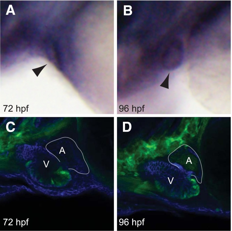
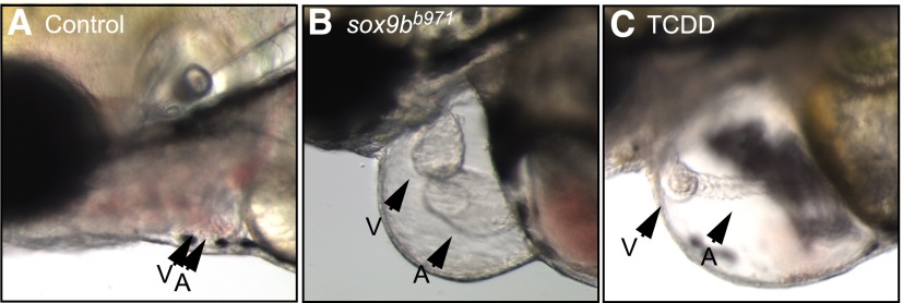
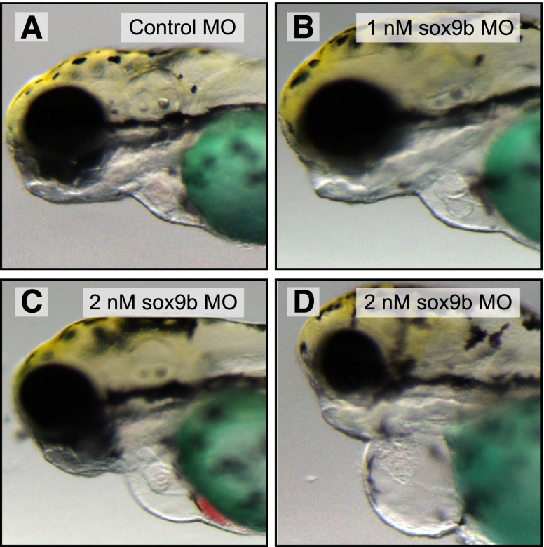
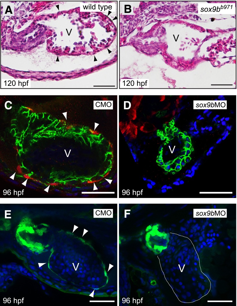
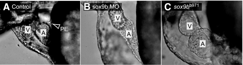
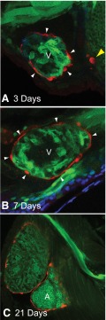
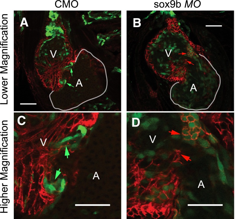
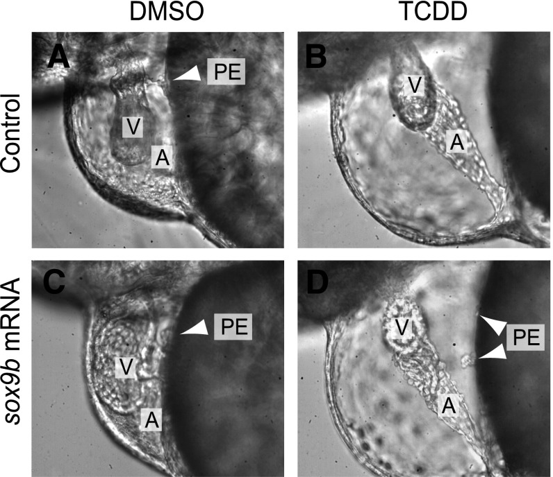
Activation of the transcription factor aryl hydrocarbon receptor by 2,3,7,8-tetrachlorodibenzo-p-dioxin (TCDD) prevents the formation of the epicardium and leads to severe heart malformations in developing zebrafish (Danio rerio). The downstream genes that cause heart malformation are not known. Because TCDD causes craniofacial malformations in zebrafish by downregulating the sox9b gene, we hypothesized that cardiotoxicity might also result from sox9b downregulation. We found that sox9b is expressed in the developing zebrafish heart ventricle and that TCDD exposure markedly reduces this expression. Furthermore, we found that manipulation of sox9b expression could phenocopy many but not all of the effects of TCDD at the heart. Loss of sox9b prevented the formation of epicardium progenitors comprising the proepicardium on the pericardial wall, and prevented the formation and migration of the epicardial layer around the heart. Zebrafish lacking sox9b showed pericardial edema, an elongated heart, and reduced blood circulation. Fish lacking sox9b failed to form valve cushions and leaflets. Sox9b is one of two mammalian Sox9 homologs, sox9b and sox9a. Knock down of sox9a expression did not cause cardiac malformations, or defects in epicardium development. We conclude that the decrease in sox9b expression in the heart caused by TCDD plays a role in many of the observed signs of cardiotoxicity. We find that while sox9b is expressed in myocardial cells, it is not normally expressed in the affected epicardial cells or progenitors. We therefore speculate that sox9b is involved in signals between the cardiomyocytes and the nascent epicardial cells.


Figures

Fig. 1.
Fig. 1.
Sox9b is expressed in the zebrafish larval heart. Wild-type AB zebrafish carrying sox9b:EGFP were collected at the indicated times for either in situ hybridization with a sox9b probe (A and B) or confocal microscopy (C and D). Lateral views are shown in all images, with head extending leftward, and the yolk sac to the right, with n = 6 fish examined per group. For the in situ images, the arrowheads point to the heart. For the confocal images, the white outline indicates the border of the atrium, indicated as A; the ventricle is indicated as V. The sox9b-GFP signal is shown in green. Blue indicates immunostaining for activated leukocyte cell adhesion molecule (ALCAM).
Fig. 2.
Fig. 2.
TCDD reduces sox9b expression in the zebrafish larval heart. Zebrafish embryos carrying the sox9b:EGFP reporter were exposed to TCDD as described in Materials and Methods. Embryos were examined at 72 hpf using confocal microscopy and representative lateral images are shown: (A) control heart, (B) TCDD-treated. White outline denotes ventricle (V); anterior is to the left. (C) Hearts were isolated at 72 hpf for quantitative reverse-transcription polymerase chain reaction measurement of sox9b expression, normalized to β-actin mRNA for each treatment group. Results are mean ± S.E., n = 3 replicate experiments. Asterisk denotes significantly different from controls (P < 0.05).
Fig. 3.
Fig. 3.
Loss of sox9b produces cardiac malformations that resemble those produced by TCDD. Representative brightfield photomicrographs of 96-hpf zebrafish. Lateral views are shown in all images, with head extending leftward, and the yolk sac to the right. Arrowheads denote location of the heart ventricle (V) and atrium (A). AB control and TCDD-treated fish are at left and right, respectively. The center panel shows a sox9bb971 homozygous mutant fish.
Fig. 4.
Fig. 4.
Graded doses of sox9b MO produce a range of cardiac malformation severity. Wild-type zebrafish embryos were injected with control and sox9b MOs and collected at 96 hpf for brightfield microscopy. Representative brightfield lateral images are shown with anterior to the left. (A) Control MO, (B) sox9b MO (1 nM), (C and D) sox9b MO (2 nM). The Fast Green used as part of the MO injection solution has colored the yolk green (n = 10 fish per treatment).
Fig. 5.
Fig. 5.
Sox9b is required for zebrafish epicardium development. (A and B) Brightfield images of hematoxylin and eosin–stained hearts from representative wild-type larva (A) and homozygous sox9bb971 null mutant larva (B) at 120 hpf. Black arrowheads indicate epicardial cells. (C and D) Embryos from the tcf21:DsRed epicardial cell reporter line were injected with the control (CMO) or sox9b MO (sox9bMO), and examined using confocal microscopy at 96 hpf for epicardium formation. Red indicates expression of tcf21, with white arrowheads indicating epicardial cells. Green indicates immunostaining for activated leukocyte cell adhesion molecule (ALCAM), marking cell boundaries, and the blue is 4′,6-diamidino-2-phenylindole (DAPI) staining, revealing nuclei. (E and F) Eggs from the pard3-GFP epicardial cell reporter line were injected with the control or sox9b MO, and examined using confocal microscopy at 96 hpf for epicardium formation. Green indicates expression of pard3 in epicardial cells which are indicated with white arrowheads. The blue is DAPI staining, revealing nuclei. For all panels representative images are shown with a minimum of n = 6 per group. Scale bar, 50 μm.
Fig. 6.
Fig. 6.
Formation of the proepicardium is dependent on sox9b expression. Representative lateral images of hearts within the pericardium of 72-hpf zebrafish. (A) Wild-type control; (B) wild-type zebrafish injected with sox9b MO; (C) homozygous sox9bb971 mutant (n = 10 per group; anterior to the left). Arrow and white outline indicate the “grape-like” PE in the wild-type control, and not found in the others. The atria and ventricles are marked as A and V, respectively.
Fig. 7.
Fig. 7.
Sox9b is not expressed in proepicardial or epicardial cells. Representative ventral-lateral images of the zebrafish heart in reporter fish expressing both sox9b:EGFP and tcf21:DsRed with anterior to the left (n = minimum of 8 per group). The top panel shows the heart at 3 days postfertilization (A), the middle shows a heart at 7 days (B), and the bottom panel shows the heart at 21 days (C). The yellow arrowhead indicates the proepicardium, while white arrowheads denote epicardial cells. The ventricle and atrium are marked as V and A, respectively.
Fig. 8.
Fig. 8.
Endocardial valve cushions fail to form following loss of sox9b. Representative 96-hpf ventral images of flk1:GFP zebrafish that were injected with sox9b MO or control MO (CMO) (n = 5 per group). Samples were immunostained with activated leukocyte cell adhesion molecule (ALCAM) in red to show cell boundaries. The ventricles and atria are marked as A and V, respectively. (A and B) 10× magnification; (C and D) 40× magnification. The green arrows indicate the valve cushions and nascent leaflets forming in the AV junction; the red arrows indicate the sites at the AV junction where valve cushions failed to form. Scale bar, 50 μm.
Fig. 9.
Fig. 9.
Sox9b mRNA injection can restore PE formation in fish treated with TCDD. (C and D) Wild-type (AB) embryos were injected with sox9b mRNA (200 pg) at the 1-4 cell stage, or left uninjected as controls (A and B). The embryos were then exposed to TCDD (B and D) or vehicle (A and C) (DMSO) as in previous experiments, as indicated. Images were collected with differential interference contrast (DIC) microscopy at 72 hpf. The A and V indicate the atrium and ventricle, respectively. Where present the PE is indicated by white arrowheads. Fish were raised in 0.003% propylthiourea in the water to inhibit pigment formation. Representative images are shown, with n = 46–73 individuals examined.

References

    1. Akiyama H, Chaboissier MC, Behringer RR, Rowitch DH, Schedl A, Epstein JA, de Crombrugghe B. (2004) Essential role of Sox9 in the pathway that controls formation of cardiac valves and septa. Proc Natl Acad Sci USA 101:6502–6507 - PMC - PubMed
    1. Andreasen EA, Spitsbergen JM, Tanguay RL, Stegeman JJ, Heideman W, Peterson RE. (2002) Tissue-specific expression of AHR2, ARNT2, and CYP1A in zebrafish embryos and larvae: effects of developmental stage and 2,3,7,8-tetrachlorodibenzo-p-dioxin exposure. Toxicol Sci 68:403–419 - PubMed
    1. Antkiewicz DS, Burns CG, Carney SA, Peterson RE, Heideman W. (2005) Heart malformation is an early response to TCDD in embryonic zebrafish. Toxicol Sci 84:368–377 - PubMed
    1. Antkiewicz DS, Peterson RE, Heideman W. (2006) Blocking expression of AHR2 and ARNT1 in zebrafish larvae protects against cardiac toxicity of 2,3,7,8-tetrachlorodibenzo-p-dioxin. Toxicol Sci 94:175–182 - PubMed
    1. Bartman T, Walsh EC, Wen KK, McKane M, Ren J, Alexander J, Rubenstein PA, Stainier DY. (2004) Early myocardial function affects endocardial cushion development in zebrafish. PLoS Biol 2:E129. - PMC - PubMed

Publication types

MeSH terms

Substances